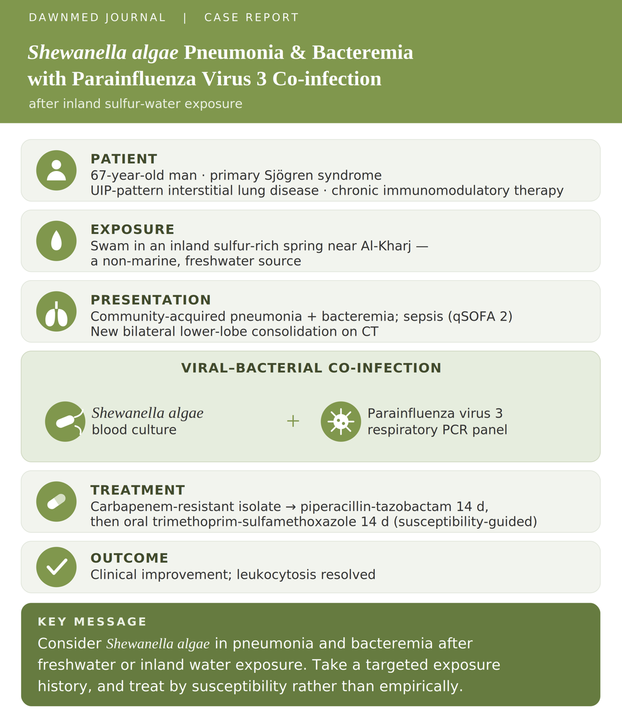
Shewanella algae Pneumonia and Bacteremia After Inland Sulfur-Water Exposure with Concurrent Parainfluenza Virus 3 Infection: A Case Report
Introduction
Shewanella algae is a halophilic, oxidase-positive, non-fermenting Gram-negative bacillus that is widely distributed in marine and other aquatic environments. Increasingly recognized as an opportunistic human pathogen, it causes infections ranging from skin and soft-tissue disease to hepatobiliary infection, bacteremia, and sepsis (1,2). Respiratory involvement remains distinctly uncommon, accounting for a small minority of reported infections and documented predominantly in isolated case reports or limited series (2,3).
Clinically significant S. algae infection occurs disproportionately in older adults and in patients with chronic comorbidities or impaired host defenses (1,2). Structural lung disease, including interstitial lung disease with traction bronchiectasis, may impair mucociliary clearance and local pulmonary defenses, while autoimmune disease and corticosteroid exposure may further increase susceptibility to invasive infection. Reported pulmonary cases have likewise involved medically vulnerable, often immunocompromised, patients (3). Infection is epidemiologically linked to occupational or recreational aquatic exposure, particularly contact with marine environments, and is reported more frequently in warmer regions (1). Although the organism is classically marine, recent whole-genome analysis of an inland S. algae bloodstream isolate found its closest genetic affinity to a freshwater Shewanella species and signatures of freshwater adaptive evolution, indicating a capacity to adapt to inland freshwater niches and supporting the plausibility of infection acquired from a non-marine inland source (4). In the present case, recent swimming in sulfur-rich spring water near Al-Kharj provided a relevant epidemiological context for the isolation of S. algae.
The interaction between S. algae and respiratory viral infection remains poorly characterized. Although bacterial coinfection has been described in adults hospitalized with human parainfluenza virus (5), and a recent report documented S. algae bacteremia associated with Epstein–Barr virus reactivation (4), to our knowledge no published case has specifically characterized concurrent S. algae respiratory infection and parainfluenza virus type 3 (PIV-3). We report a 67-year-old man with Sjögren-associated interstitial lung disease and chronic corticosteroid exposure who developed community-acquired pneumonia with S. algae bacteremia after swimming in sulfur-rich water near Al-Kharj, Saudi Arabia, with simultaneous detection of PIV-3. The case shows why an aquatic exposure history matters in this setting and points to a possible contribution of viral–bacterial coinfection in a structurally and immunologically vulnerable host.
Case Presentation
History
A 67-year-old man presented to the emergency department with a one-day history of sudden-onset productive cough yielding yellowish-green sputum, accompanied by high-grade fever documented at home up to 39 °C, rigors, and generalized fatigue. There were no preceding upper-respiratory symptoms and no known contact with unwell individuals.
His background was notable for chronic immunomodulatory therapy and structural lung disease. He had primary Sjögren syndrome maintained on hydroxychloroquine 400 mg once daily and prednisolone 5 mg on alternate days, interstitial lung disease (ILD) treated with an inhaled fluticasone furoate/vilanterol combination, and bilateral knee osteoarthritis managed with a topical diclofenac patch and tramadol. He had no significant surgical or family history.
A complete systems review was otherwise unremarkable. He specifically denied weight loss, night sweats, or anorexia; chest pain, palpitations, syncope, orthopnea, paroxysmal nocturnal dyspnea, or hemoptysis; abdominal pain, nausea, vomiting, altered bowel habit, hematemesis, or melena; urinary symptoms; headache, dizziness, altered cognition, focal neurological deficit, or neck stiffness; visual symptoms; sore throat, hoarseness, or dysphagia; arthralgia, joint swelling, or skin rash; and heat or cold intolerance, abnormal pigmentation, hair loss, or easy bruising.
Examination
On arrival the patient was conscious, alert, oriented, cooperative, and not in obvious distress. He was febrile, tachycardic, tachypneic, and hypotensive, maintaining adequate peripheral oxygen saturation on 1 L/min of supplemental oxygen via nasal cannula. Body mass index was 29 kg/m².
Across serial observations, systolic blood pressure ranged from 82 to 130 mmHg and diastolic from 47 to 60 mmHg (mean arterial pressure 55–74 mmHg), heart rate rose to 131 beats/min, respiratory rate to 35 breaths/min, and temperature peaked at 38.4 °C in the department (39 °C at home). Peripheral oxygen saturation was generally maintained in the low-to-mid 90s on 1 L/min, with a transient nadir of 75%.
Respiratory examination revealed bilateral mid-to-lower zone coarse crackles. Heart sounds were normal with a regular pulse and no murmur. There was no palpable peripheral lymphadenopathy, no joint swelling, tenderness, rash, or discoloration, and no focal neurological deficit. The abdomen was soft, lax, and non-tender with a normal contour, and there was no bilateral lower-limb pitting edema.
Investigations
Laboratory studies demonstrated a neutrophil-predominant leukocytosis with markedly raised inflammatory markers. Serum sodium was mildly reduced, renal and hepatic function were preserved, and there was mild hypophosphatemia, borderline-low magnesium, and a mildly elevated lactate. Random glucose was mildly elevated at 9.9 mmol/L, with heavy glucosuria (≥1000 mg/dL) and ketonuria; the degree of glucosuria was disproportionate to the concurrent serum glucose, which lies close to the usual renal threshold, raising the possibility of transient hyperglycemia, a low renal glucose threshold, or unrecognized dysglycemia rather than a simple corticosteroid effect. Cardiac biomarkers were within normal limits, and a blood gas showed a mild respiratory alkalosis in keeping with the tachypnea. Full results are summarized in Tables 1a–1c.
Table 1a. Hematology and inflammatory markers
| Investigation | Result | Reference (adult) |
|---|
| White cell count | 16.10 → 9.23 ×10⁹/L | 4.0–11.0 |
| Neutrophils | 79.2% → 54.8% (ANC 12.70 → 5.06 ×10⁹/L) | 40–75% |
| Lymphocytes | 8.34% → 30.0% (abs. 1.34 ×10⁹/L) | 20–45% |
| Hemoglobin | 141 → 135 g/L | 130–170 (M) |
| Red cell count | 4.82 → 4.63 ×10¹²/L | 4.5–5.9 |
| C-reactive protein | 89.90 mg/L | <5 |
| Procalcitonin | 1.08 ng/mL | <0.5 |
| ESR | 60 mm/hr | <15 |
Table 1b. Chemistry and metabolic profile
| Investigation | Result | Reference (adult) |
|---|
| Sodium | 131 mmol/L | 135–145 |
| Potassium | 4.2 mmol/L | 3.5–5.1 |
| Chloride | 96 mmol/L | 98–107 |
| Bicarbonate | 24 mmol/L | 22–29 |
| Urea (BUN) | 4.1 mmol/L | 2.5–7.1 |
| Creatinine | 60 µmol/L | 60–110 (M) |
| eGFR | 124 mL/min/1.73m² | >90 |
| Total protein | 82 g/L | 60–80 |
| Albumin | 42 g/L | 35–50 |
| Total bilirubin | 15.4 µmol/L | <21 |
| Alkaline phosphatase | 107 U/L | 40–130 |
| ALT / AST | 16 / 19 U/L | <41 / <40 |
| Calcium (adjusted) | 2.25 (2.21) mmol/L | 2.20–2.60 |
| Magnesium | 0.66 mmol/L | 0.70–1.00 |
| Phosphate | 0.65 mmol/L | 0.80–1.50 |
| Random glucose | 9.9 mmol/L | <11.1 |
| Lactate | 2.12 mmol/L | 0.5–2.0 |
| Creatine kinase | 85 U/L | 39–308 |
| Anion gap | 15 | 8–16 |
Table 1c. Coagulation, cardiac, and blood-gas indices
| Investigation | Result | Reference (adult) |
|---|
| PT / INR / aPTT | 11.8 s / 1.09 / 26.2 s | 11–14 / 0.8–1.2 / 25–35 |
| Troponin I | 18.2 ng/L | assay-dependent |
| BNP | 24.4 pg/mL | <100 |
| pH | 7.47 | 7.35–7.45 |
| pCO₂ / pO₂ | 38 / 48 mmHg | 35–45 / 80–100 |
| HCO₃⁻ / base excess | 27.7 mmol/L / +4.0 | 22–26 / −2 to +2 |
| SatO₂ / O₂Hb / COHb / MetHb | 81.6% / 79.6% / 2.1% / 0.3% | — |
Reference intervals are typical adult values and may vary by laboratory. Paired values (X → Y) denote admission and repeat results.
Urinalysis showed clear, straw-colored urine (specific gravity 1.015, pH 6.5) with glucose ≥1000 mg/dL and ketones; blood, leukocyte esterase, nitrite, protein, and bilirubin were negative, urobilinogen was normal, and mucus was rare.
A multiplex respiratory pathogen PCR panel detected parainfluenza virus 3. All other targets were not detected, including influenza A and B, respiratory syncytial virus, SARS-CoV-2, MERS-CoV, adenovirus, rhinovirus/enterovirus, human metapneumovirus, the remaining parainfluenza (1, 2, 4) and coronavirus (NL63, 229E, OC43, HKU1) subtypes, Mycoplasma pneumoniae, Chlamydia pneumoniae, Bordetella pertussis, and Bordetella parapertussis.
A 12-lead electrocardiogram showed sinus tachycardia up to 131 bpm without ischemic ST–T changes.
Imaging
A portable chest radiograph demonstrated new bilateral lower-zone infiltrative changes and patchy airspace opacities superimposed on chronic interstitial markings, compared with a prior film.
CT pulmonary angiography (CTPA) excluded acute pulmonary embolism: the pulmonary trunk and main, segmental, and subsegmental pulmonary arteries opacified without filling defect, the pulmonary trunk measured 2.6 cm, and there was no right-heart strain, pulmonary hypertension, or pleural or pericardial effusion. On a background of established usual interstitial pneumonia (UIP)-pattern ILD, comprising bilateral basilar-predominant subpleural reticulation, interlobular septal thickening, traction bronchiectasis, and scattered pulmonary nodules, there was interval development of bilateral lower-lobe consolidation and ground-glass opacity, more marked on the right, in keeping with a superimposed infective process. Incidental findings included severe coronary artery and right pericardial calcification, multiple reactive-appearing mediastinal and hilar lymph nodes (largest 1.3 cm, right paratracheal), a centrally patent tracheobronchial tree, and an interval severe L1 compression fracture with minimal retropulsion on a background of spondylotic change and osteopenia. The radiological impression was known Sjögren disease with UIP-pattern ILD complicated by new bilateral lower-lobe opacities suggestive of active infection; no pulmonary embolism; with follow-up imaging advised in 6–8 weeks.
Microbiology and exposure history
Blood cultures grew a Gram-negative bacillus, subsequently identified as Shewanella algae. Antimicrobial susceptibility testing (E-test) is shown in Table 2, notable for carbapenem resistance with retained susceptibility to trimethoprim-sulfamethoxazole, ciprofloxacin, and ceftazidime.
Table 2. Antimicrobial susceptibility of the blood-culture isolate (E-test)
| Antimicrobial | MIC (µg/mL) | Interpretation |
|---|
| Trimethoprim-sulfamethoxazole | 0.25 | Susceptible |
| Ciprofloxacin | 0.19 | Susceptible |
| Ceftazidime | 0.19 | Susceptible |
| Piperacillin-tazobactam | 24 | Intermediate |
| Meropenem | 8 | Resistant |
Because Shewanella is an uncommon, aquatic-associated organism, the infectious diseases team revisited the history and elicited that the patient had recently been swimming in naturally occurring sulfur-rich (hot spring) water near Al-Kharj, Saudi Arabia, establishing a plausible environmental source for an opportunistic Shewanella algae infection in a susceptible host.
Management and outcome
The patient was admitted under the pulmonology service as an ILD exacerbation secondary to community-acquired pneumonia and commenced empirically on intravenous ceftriaxone and oral azithromycin. On presentation he met criteria for sepsis (qSOFA 2, from a systolic blood pressure below 100 mmHg and a respiratory rate of 35/min with preserved mentation, in the context of infection, hypotension, and a lactate of 2.12 mmol/L); he was managed on the pulmonology ward, and his blood pressure subsequently recovered (systolic up to 130 mmHg). After Shewanella algae was isolated from blood and the infectious diseases team had reviewed susceptibilities and exposure history, therapy was changed to intravenous piperacillin-tazobactam for a total of 14 days, followed by an oral step-down course of trimethoprim-sulfamethoxazole for a further 14 days. Although the isolate tested intermediate to piperacillin-tazobactam by E-test, while ceftazidime and ciprofloxacin were fully susceptible (MIC 0.19 each), the patient was already improving clinically on piperacillin-tazobactam; the infectious diseases team elected to continue it with dose optimization to achieve adequate exposure against an intermediate isolate rather than change an effective regimen, before stepping down to oral trimethoprim-sulfamethoxazole, to which the isolate was fully susceptible.
The patient improved on treatment, with normalization of the leukocytosis on repeat testing during admission (white cell count 9.23 ×10⁹/L, neutrophils 54.8%) and a corresponding rise in the lymphocyte fraction. Interval chest imaging was planned at 6–8 weeks to confirm radiological resolution.
Table 3. Clinical timeline
| Time point | Event |
|---|
| Day 0 (presentation) | One-day productive cough, fever to 39 °C, rigors, and fatigue; hypotensive, febrile, tachycardic, and tachypneic on 1 L/min oxygen; met criteria for sepsis (qSOFA 2). |
| Initial workup | Neutrophilic leukocytosis (WBC 16.10, ANC 12.70), CRP 89.90, PCT 1.08, ESR 60, lactate 2.12; chest radiograph with new bilateral infiltrates. |
| Imaging | CTPA excluded pulmonary embolism; UIP-pattern ILD with new bilateral lower-lobe consolidation/ground-glass opacity. |
| Virology | Respiratory panel positive for parainfluenza virus 3; other pathogens not detected. |
| Empirical therapy | Admitted under pulmonology; started on ceftriaxone plus azithromycin. |
| Microbiology | Blood culture grew Shewanella algae (carbapenem-resistant; susceptible to TMP-SMX, ciprofloxacin, ceftazidime). |
| Exposure history | Recent swimming in sulfur-rich spring water near Al-Kharj identified by the ID team. |
| Definitive plan | Changed to piperacillin-tazobactam for 14 days, then oral trimethoprim-sulfamethoxazole for 14 days. |
| Follow-up | Leukocytosis normalized during admission; interval CT advised at 6–8 weeks. |
Discussion
Three features of this case are unusual: Shewanella algae is a rare cause of pneumonia with bacteremia; the organism was acquired from an inland sulfur-rich spring rather than the marine or coastal environment classically described; and it occurred together with parainfluenza virus type 3 (PIV-3), a viral–bacterial coinfection, in a host with structural lung disease and chronic immunomodulatory therapy (2,3).
S. algae is an oxidase-positive, motile, non-fermenting Gram-negative bacillus and is the species responsible for most human Shewanella infections (1,6). Human infection correlates with the temperature and salinity of seawater, which explains the predominance of cases in warm climates and warm seasons, a pattern relevant to the climate of Saudi Arabia (6). The organism's environmental adaptability and virulence-associated features have increasingly been characterized at the genomic level (1,4).
Shewanella infection is epidemiologically linked to occupational or recreational aquatic exposure, classically marine or estuarine contact, and to consumption of raw seafood (1,6). The risk of water-associated infection varies by water type, and contaminated natural water sources are recognized to harbor unusual organisms with atypical resistance profiles, warranting a directed diagnostic and therapeutic approach (7). In this patient, the only identified exposure was swimming in a sulfur-rich inland spring near Al-Kharj, a non-marine source. Although S. algae is classically halophilic and marine-associated, the inland bloodstream isolate characterized by whole-genome analysis showed its closest affinity to a freshwater Shewanella species and signatures of freshwater adaptive evolution, supporting the plausibility of acquisition from inland spring or mineral water (4). This exposure came to light only after the organism was isolated, which argues for taking a targeted environmental history whenever an unusual aquatic organism is grown.
Invasive S. algae infection occurs almost exclusively in older adults and in hosts with chronic comorbidity or immune compromise; in the largest case series, infection was strongly associated with underlying hepatobiliary disease and malignancy, and primary bacteremia may follow a fulminant course in immunocompromised patients (2,6). Our patient carried several compounding susceptibilities: advanced age, Sjögren syndrome, long-term hydroxychloroquine and low-dose alternate-day corticosteroid therapy, and usual interstitial pneumonia-pattern interstitial lung disease with traction bronchiectasis. His immunomodulation was modest rather than profound, and the term immunocompromised should be applied with caution; nonetheless, autoimmune disease, chronic corticosteroid exposure, mild lymphopenia (absolute lymphocyte count 1.34 ×10⁹/L), and structural lung disease that impairs mucociliary clearance may together have lowered his threshold for invasive infection.
Pulmonary involvement is among the least common Shewanella syndromes, with the prior literature limited to isolated reports such as S. algae pneumonia with bacteremia in an elderly long-term-care resident (2,3). The contribution of antecedent or concurrent viral infection to bacterial pneumonia is well recognized, and PIV is an established cause of lower respiratory tract infection in hospitalized adults, among whom bacterial coinfection has been described (5). In the present case, PIV-3 was the only respiratory virus detected, and viral injury to the respiratory epithelium may have facilitated secondary bacterial invasion; an analogous synergistic interaction between S. algae bloodstream infection and a reactivated herpesvirus (Epstein–Barr virus) has recently been reported to accelerate septic shock (4). To our knowledge, concurrent S. algae respiratory infection and PIV-3 has not previously been described.
The isolate was resistant to meropenem and intermediate to piperacillin-tazobactam, while remaining susceptible to ceftazidime, ciprofloxacin, and trimethoprim-sulfamethoxazole. Carbapenem resistance among Shewanella has risen, and the genus is recognized as an environmental reservoir and probable progenitor of antibiotic-resistance genes, including chromosomally encoded β-lactamase and quinolone-resistance determinants (2,8). Reported therapeutic options for S. algae include third- and fourth-generation cephalosporins, fluoroquinolones, aminoglycosides, piperacillin, carbapenems, and trimethoprim-sulfamethoxazole, although susceptibility is variable and treatment should be guided by testing (1). In this patient, the intermediate agent was continued with dose optimization on the basis of clinical response, with an oral step-down to fully susceptible trimethoprim-sulfamethoxazole.
Two practical points follow. First, a detailed water and environmental exposure history is worth taking when an unusual aquatic organism is isolated. Second, early infectious-diseases involvement changed management here: it redirected the history, identified the likely source, and guided antimicrobial therapy.
Several limitations should be acknowledged. As a single case, causal inferences are limited. The environmental source was inferred from history; the implicated water was not cultured, so a definitive microbiological link could not be established. The relative pathogenic contributions of PIV-3 and S. algae cannot be disentangled, and respiratory specimens for bacterial culture were not available to confirm pulmonary involvement by the same organism. We also acknowledge a tension in antimicrobial management: although we emphasize susceptibility-guided therapy, piperacillin-tazobactam (intermediate) was continued when ceftazidime and ciprofloxacin tested fully susceptible, a choice that reflected clinical improvement on the existing regimen and was followed by an oral step-down to a fully susceptible agent. Finally, longer-term clinical and radiological outcomes were limited, with follow-up imaging planned but not yet available at the time of reporting.
Conclusion
This case shows that Shewanella algae can cause severe respiratory infection with bacteremia outside its classically described marine setting, here following exposure to an inland sulfur-rich spring. Concurrent detection of parainfluenza virus type 3 raises the possibility of viral–bacterial synergy in a host rendered vulnerable by structural lung disease and chronic immunomodulatory therapy. The isolate's carbapenem resistance, despite retained susceptibility to other agents, argues for susceptibility-guided rather than empirical therapy when an atypical aquatic organism is identified. Clinically, it reinforces the value of a directed environmental exposure history in patients with unexplained pneumonia and bacteremia, particularly in those with structural lung disease or impaired host defenses. As an isolated observation, it cannot establish causality between exposure, viral coinfection, and disease severity; further reports characterizing inland-acquired Shewanella infections and their interaction with respiratory viruses are needed to clarify risk and guide management.
Declarations
Ethics Approval: Not applicable.
Consent for Publication: Written informed consent was obtained from the patient for publication of this case report.
Availability of Data and Materials: All data are included in this article. Further information is available from the corresponding author on reasonable request.
Competing Interests: The authors declare no competing interests.
Funding: None.
Author Contributions: All authors contributed to the conception of the report, acquisition and interpretation of the clinical data, and drafting and critical revision of the manuscript. All authors read and approved the final manuscript and agree to be accountable for all aspects of the work.
Acknowledgements: None.