Minimally Invasive Surgery for Ankle and Distal Tibial Fractures: A Narrative Review
Introduction
Scope and review approach
Ankle fractures and distal tibial fractures near the ankle present related but distinct reconstructive problems. This narrative review examines minimally invasive surgery for unstable ankle fractures, distal-fibular fractures, extra-articular distal tibial metaphyseal fractures, and intra-articular pilon fractures. It is a narrative rather than a systematic review. Pilon fractures remain in scope because they share the problems of an injured soft-tissue envelope and ankle-joint reconstruction, but they are considered separately from extra-articular distal tibial fractures. The techniques addressed are minimally invasive plate osteosynthesis (MIPO) of the distal fibula, MIPO of the distal tibia, and fibular intramedullary nailing.
Epidemiology and injury spectrum
Elsoe and colleagues examined 9,767 ankle fractures treated during 2005–2014 [1]. They reported a mean incidence of 168.7 per 100,000 people per year [1]. Lateral-malleolar fractures represented 55% of the cohort [1]. Falls accounted for 61% of injuries and sports for 22% [1]. Gundtoft and colleagues subsequently identified 155,740 ankle fractures in a nationwide Danish cohort [2]. The mean incidence during 1997–2018 was 164 per 100,000 person-years [2]. Incidence increased over that period, principally among women and older patients [2]. The regional and nationwide estimates describe different source populations and should not be treated as interchangeable.
The term “ankle fracture” covers injuries with different combinations of malleolar, ligamentous, syndesmotic, and articular disruption. A fixation method suited to an isolated distal-fibular pattern cannot be transferred automatically to a comminuted tibial plafond injury.
Mortise congruence and the rationale for fixation
Operative reduction seeks to restore the relationship between the talus and the ankle mortise. In a cadaveric experiment, Ramsey and Hamilton found that the initial millimetre of lateral talar displacement reduced tibiotalar contact area by an average of 42% [3]. Lloyd and colleagues repeated the experiment in another cadaveric model and found a 40% loss after one millimetre of lateral shift, with substantial variation related to ankle morphology [4]. The replication supports the biomechanical observation, but neither experiment establishes a clinical outcome threshold for surgery or post-traumatic arthritis.
Open reduction and internal fixation (ORIF) permits direct manipulation of the fracture and plate placement. Kurt and colleagues described ORIF as a long-established technique for lateral-malleolar fractures [5]. In their MIPO protocol, the adequacy of fracture reduction was assessed under fluoroscopic guidance [5]. Minimally invasive fixation does not change the objective of restoring length, rotation, mortise alignment, and any articular injury. It changes the exposure and the means used to obtain and verify reduction.
Techniques within the review
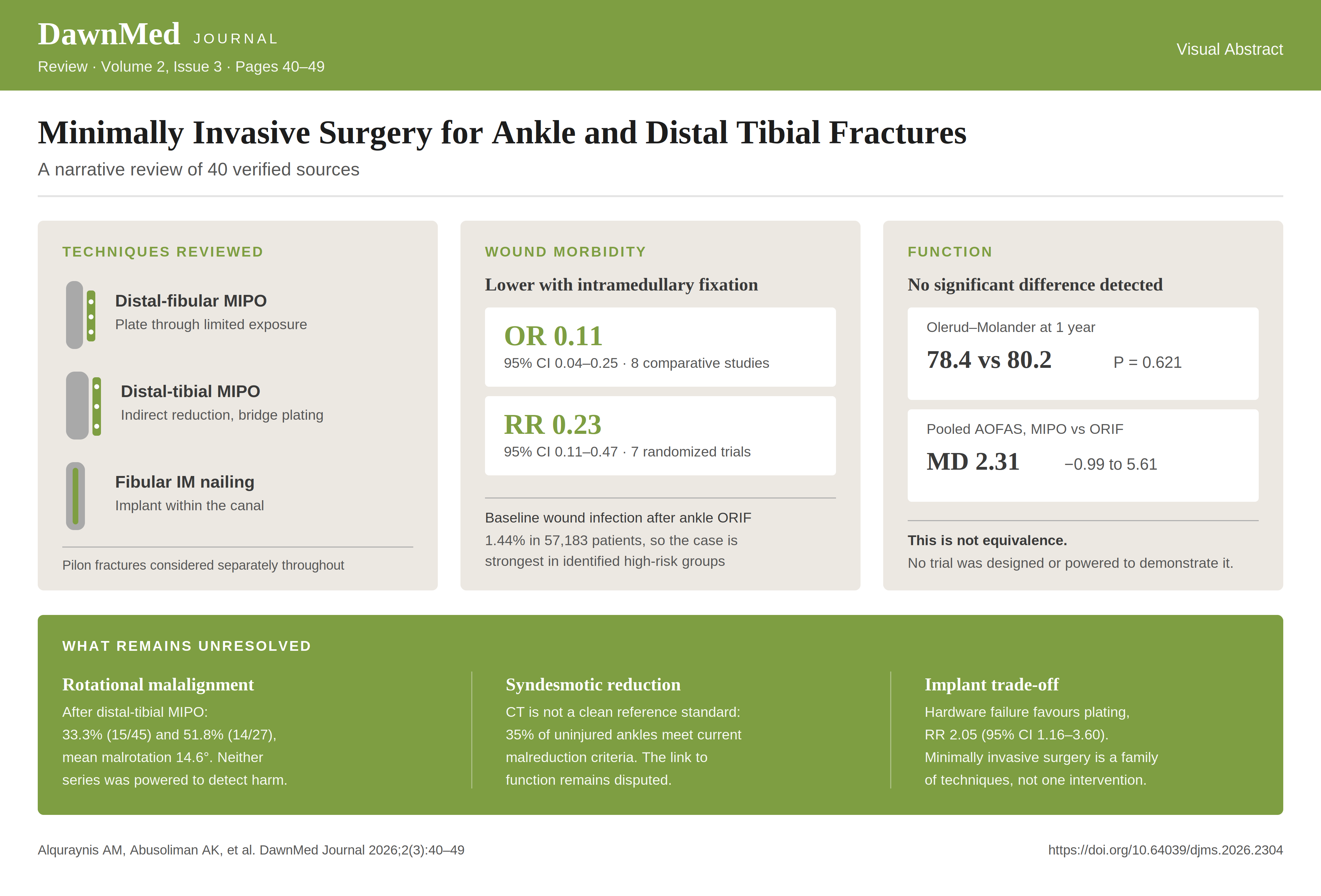
The label “minimally invasive” includes operations with different mechanical and biological compromises. Distal-fibular MIPO retains plate fixation while limiting exposure at the fracture. Distal-tibial MIPO uses indirect reduction and bridge plating across the metaphysis. Fibular intramedullary nailing places the principal implant within the bone through a limited approach. These methods are examined separately because access, reduction control, implant position, and syndesmotic fixation differ among them.
The soft-tissue problem
At population level, SooHoo and colleagues characterized the overall short-term complication rate after ankle ORIF as low [6]. Their California discharge database included 57,183 inpatient ORIF cases [6]. Wound infection, captured through readmission within 90 days, occurred in 1.44% [6]. This population estimate does not make the average patient the main soft-tissue case for minimally invasive fixation. The rationale is stronger in patients and injuries with identifiable wound risk. Miller and colleagues associated wound complications with diabetes, peripheral neuropathy, open fracture, wound-compromising medication, and postoperative nonadherence [7]. At the distal tibia, limited soft-tissue coverage and compromised blood supply add to the difficulty of treatment [8]. Lau and colleagues nevertheless documented late wound infection and implant impingement after distal-tibial MIPO [9]. A smaller approach may reduce surgical exposure, but it does not remove soft-tissue or implant-related risk.
Distal tibial fracture categories must remain separate when these trade-offs are assessed. Extra-articular metaphyseal fractures do not require reconstruction of the tibial plafond. Pilon fractures are intra-articular distal tibial injuries, and Daniels and colleagues described the combined difficulty created by high-energy trauma, articular comminution, and soft-tissue damage [10]. Sirkin and colleagues described initial ankle-spanning external fixation followed by definitive articular reconstruction after soft-tissue swelling had subsided [11]. That staged protocol supplies context for soft-tissue management in pilon fractures; it is not a direct comparison of MIPO with ORIF.
This review therefore keeps distal-fibular MIPO, distal-tibial MIPO, fibular nailing, staged fixation, and external fixation analytically distinct. It reports study-specific populations, denominators, and outcome definitions rather than combining unlike endpoints into pooled narrative ranges.
Techniques
Terminology and selection
“Minimally invasive” describes an access strategy, not a single operation. Distal-fibular MIPO retains an extramedullary plate but limits exposure of the fracture and plate track [5,12,13]. Distal-tibial MIPO uses indirect reduction and a plate passed along the bone through separated incisions [14]. Fibular intramedullary nailing moves the main implant into the medullary canal while still requiring reduction, distal entry, locking, and assessment of the syndesmosis [15,16]. These operations should therefore be compared by fracture location, reduction method, implant, and treatment of associated malleolar or syndesmotic injury.
Selection begins with whether length, rotation, ankle-mortise alignment, and any articular displacement can be restored and verified through the intended access. A limited approach can be enlarged when indirect or percutaneous manoeuvres do not produce an acceptable reduction. Soft-tissue compromise may make a smaller approach attractive, but it does not make a mechanically unsuitable fracture suitable for that implant. The published protocols also differ: some use closed reduction, some use a short incision directly over the fracture, and some combine percutaneous plate or nail insertion with open treatment of another component [12,14,16]. “MIPO” should not be read as “closed fixation.”
Distal-fibular MIPO
The distal-fibular MIPO construct is a plate placed through a limited lateral incision. In the Marazzi protocol, a 2-cm incision was made distal to the malleolar tip and the plate was advanced subcutaneously in a retrograde direction [12]. Fracture reduction was then checked fluoroscopically [12]. Reduction may occur as the contoured plate is seated, or it may require percutaneous clamps. Screws are inserted through the distal wound and small proximal incisions. The fracture is not routinely exposed along the full plate length.
Kurt and colleagues used a related limited-access method in a randomized comparison with ORIF [5]. Their protocol retained the fracture haematoma, used closed reduction, and placed planned screws through mini-incisions [5]. Fluoroscopy replaces part of the direct visual information available during ORIF, so the surgeon must verify the mortise and the fibular relationship to the distal tibia before completing fixation. If the reduction remains uncertain, a short direct fracture incision is compatible with a minimally invasive strategy and may be safer than accepting an indirect reduction that cannot be confirmed.
Yañez Arauz reported a prospective series of 451 displaced AO type B ankle fractures treated with minimally invasive surgery [13]. The large single-arm experience establishes that the method can be applied repeatedly to a defined trans-syndesmotic fracture population, but it does not establish a comparative indication or prove that the same method is appropriate for comminuted, supra-syndesmotic, or pilon-associated fractures [13].
Distal-tibial MIPO
Extra-articular distal-tibial MIPO is usually a bridge-plating operation. Traction, clamps, temporary wires, or a reduction device restore length and coronal, sagittal, and rotational alignment before a precontoured plate is passed along the metaphysis. Akdemir and colleagues performed reduction under fluoroscopy, advanced a medial distal-tibial plate over the periosteum, and used a distal incision with a separate proximal mini-incision for screw placement [14]. This preserves the limited plate track but gives less direct access to the fracture than ORIF.
Intra-articular pilon fractures require a separate account. The articular surface must be reconstructed; minimally invasive plate insertion does not remove that requirement. In the Akdemir protocol, joint separation was treated with percutaneous cannulated screws before plate fixation [14]. Other pilon patterns require direct articular exposure, and the plate may then be inserted through a limited corridor after the joint block has been reconstructed. The operation should be described by both components—articular reduction and metaphyseal fixation—rather than labelled simply as MIPO.
Erivan and colleagues randomized 54 pilon-fracture patients to MIPO or ORIF using distal-tibial locking plates [17]. That trial confirms that a MIPO-versus-ORIF comparison has been attempted in an intra-articular population [17]; it does not justify transferring extra-articular results to pilon fractures. Staged external fixation remains a separate soft-tissue strategy for selected high-energy injuries [11]. Timing, temporary stabilization, articular access, and final plate insertion are distinct decisions.
Fibular intramedullary nailing and syndesmotic fixation
A fibular nail is inserted from the distal fibula after the fracture has been reduced. Modern constructs add distal and proximal locking; some permit trans-syndesmotic fixation through the implant [15,16]. The small entry wound reduces implant exposure beneath the lateral skin, but nail passage does not itself restore fibular length or rotation. Reduction may be closed, percutaneous, or mini-open.
White and colleagues randomized 125 adults to nail or plate fixation, illustrating use of a locked nail in unstable ankle fractures [15]. Ricketts and colleagues later described a minimally invasive open technique that directly reduced the fibular fracture and syndesmosis before locked nail fixation in 150 consecutive patients [16]. Because the latter was a single-surgeon case series without a plate control, its technique and results should not be treated as comparative evidence [16].
Syndesmotic fixation can be delivered through a limited corridor, but implant delivery and reduction accuracy are separate questions. In a matched cadaver experiment using 14 donors and 28 limbs, Shaner and colleagues found more internal-rotation malreduction with fluoroscopy-only minimally invasive reduction than with open reduction plus fluoroscopy [18]. Bastías and colleagues obtained immediate bilateral CT after third-generation fibular nailing in 28 patients and found a difference in fibular translation relative to the uninjured ankle (P=.01) [19]. Because it was a prospective cohort of nail-treated patients, it did not estimate a nail-versus-plate effect [19]. Nail-based fixation therefore does not remove the need to reduce and verify the syndesmosis.
Functional and radiological outcomes
Outcome framework
The evidence is easier to interpret when outcomes are kept separate. Patient-reported function, clinician-rated ankle scores, pain, return to activity, alignment, reduction quality, time to union, union status, infection, implant failure, implant removal, and unplanned reoperation answer different questions. A high union proportion cannot substitute for a functional score, and elective implant removal cannot be merged with reoperation for infection or loss of reduction. Denominators also change when only part of a cohort reaches a given follow-up.
Radiological assessment also differs by location. Distal-fibular studies usually describe ankle-mortise measures and fibular position, whereas distal-tibial studies may report coronal and sagittal distal-tibial angles. A postoperative measurement can describe alignment without establishing how much deviation changes symptoms or later arthritis. Reduction quality, alignment, and patient function should therefore appear as linked but separate results.
Comparative studies require equally careful wording. Failure to detect a statistically significant difference does not establish equivalence, especially in small trials. The sections below therefore state that no significant difference was detected when that is what the analysis showed. They do not recast those analyses as proof that two methods are interchangeable.
Distal-fibular MIPO versus ORIF
Kurt and colleagues randomized 68 patients, with 34 assigned to each technique [5]. Early postoperative pain was lower in the MIPO group, but no significant difference was detected in AOFAS scores at the reported later assessments [5]. The result describes an early recovery difference without proof of a long-term functional advantage. The trial excluded bimalleolar and trimalleolar fractures requiring fixation [5], which narrows its application to more limited lateral-malleolar injury patterns.
Marazzi and colleagues retrospectively compared 35 MIPO cases with 35 ORIF cases [12]. The study reported an overall complication rate of 14% after MIPO and 37% after ORIF (P=.029) [12]. Its individual wound, infection, nonunion, and pain comparisons were not statistically significant, so the total-complication result should not be converted into separate claims of superiority for those component endpoints [12]. This was a single-centre retrospective series, and treatment allocation was not randomized [12].
Yañez Arauz followed 451 displaced AO type B fractures treated with MIPO for a mean of 112 months [13]. The abstract reports a mean long-term AOFAS score of 97.5 and a postoperative complication incidence of 2.7% [13]. These values belong to one prospective single-arm series [13]. They do not supply an ORIF comparison, and the complication figure should not be combined with infection, implant-removal, or reoperation endpoints from other studies.
The absence of a common patient-reported instrument across these reports limits synthesis [5,12,13]. AOFAS values combine patient symptoms and examiner findings, while pain scores and return-to-activity measures capture different parts of recovery. A difference detected in the first postoperative weeks may disappear at later assessment without showing that the early difference was unimportant. Conversely, a favourable long-term score in a single-arm series does not show that another fixation method would have produced a worse result.
Together, these studies show that distal-fibular MIPO can preserve plate fixation while limiting exposure. The comparative evidence remains thinner than the size of the largest single-arm series might imply [5,12,13]. Fracture selection, associated malleolar injury, reduction method, and outcome definition differ across the reports.
Fibular nail versus plate fixation
The randomized trials do not support a single effect estimate that applies to every nail design and patient group [15,20,21]. Asloum and colleagues randomized 71 patients, but intramedullary fixation was technically impossible in seven and those cases were converted to plating [20]. The final comparative analysis contained 60 patients [20]. The nail group had fewer reported complications and higher functional scores, but the authors restricted their conclusion to non-comminuted fractures without syndesmotic injury [20]. The 7% versus 56% complication comparison was per-protocol rather than intention-to-treat: the seven technically unsuccessful nail procedures had been converted to plating and were excluded before complications were counted [20]. Excluding those technical failures from the nail arm biases the comparison in favour of nailing [20]. The conversions are part of the result because canal size, comminution, and unresolved diastasis affect whether a proposed nail construct can be completed.
White and colleagues randomized 100 patients older than 65 years [21]. Wound infection was less frequent after nailing (P=.002), while no significant difference was detected in the one-year Olerud–Molander score (63 after nailing and 61 after ORIF; P=.61) [21]. A later trial by White and colleagues randomized 125 adults younger than 65 years [15]. At one year, mean Olerud–Molander scores were 78.4 after nailing and 80.2 after plating (P=.621); no significant difference was detected [15]. Wound infections occurred in two nail patients and nine plate patients, but that comparison also did not reach statistical significance [15]. The two trials therefore produced different statistical findings for wound infection in different age-defined populations [15,21].
Tas and colleagues published a systematic review and meta-analysis, not a retrospective cohort [22]. Their review included 26 studies and 1,710 patients, while the quantitative comparison used eight comparative studies [22]. Intramedullary fixation was associated with fewer wound-related complications (OR 0.11, 95% CI 0.04–0.25; P<.01) [22]. That estimate should be read with its mixture of randomized and observational designs, devices, and populations [22].
Jain and colleagues had earlier assembled 17 intramedullary-fixation studies comprising 1,008 patients [23]. Heterogeneity in study populations, devices, and outcome reporting prevented an accurate meta-analysis [23]. Its participant total is descriptive and should not be added to the Tas total as if it were a separate comparative cohort [22,23].
Review chronology explains part of the overlap. Jain summarized early nail experience; Tas added a comparative meta-analysis that included observational evidence; Zhang, Luo, and Chen later restricted synthesis to randomized trials [22,23,24]. Each review asks a narrower or differently framed question, but later publication does not make the patients in earlier primary trials new participants [22,23,24]. Device generations also changed, so a pooled label such as intramedullary fixation can conceal compression screws, unlocked nails, and locked nails with different failure modes.
The RCT-only synthesis by Zhang, Luo, and Chen included seven trials and 586 participants [24]. No significant functional difference was detected at the reported follow-up intervals [24]. The pooled infection result favoured the nail (RR 0.23, 95% CI 0.11–0.47; P<.0001), whereas hardware failure favoured plating (RR 2.05 for nail versus plate, 95% CI 1.16–3.60; P=.01) [24]. Its included-study table contains Asloum 2014, White 2016, and White 2022 [24]. These trials also sit within the broader body evaluated by the earlier reviews. The totals of 1,710, 1,008, and 586 participants are therefore overlapping descriptions of the evidence base, not independent samples, and they must not be summed [22,23,24].
Ricketts and colleagues add a newer locked-nail case series rather than a plate comparison [16]. Of 150 consecutive patients, 113 were followed to clinical and radiographic union; 110 united after the index procedure, giving 97.3% within that followed subset [16]. The abstract reports nine unplanned reoperations and labels this 8% [16]. Because nine of 113 is approximately 8%, whereas nine of 150 is 6%, this review infers that the followed cohort was the denominator; the abstract does not print that denominator beside the reoperation statement [16]. The abstract also reports a mean PROMIS Physical Function score of 37.5 [16]. It should not be described as favourable; the abstract provides no comparator or threshold that would support that adjective [16].
Distal-tibial outcomes
Akdemir and colleagues retrospectively compared 20 MIPO cases with 24 ORIF cases [14]. No significant difference was detected in postoperative AOFAS score or the measured anterior and lateral distal-tibial angles [14]. Union time and nonunion also did not differ significantly [14]. These findings do not prove equivalence.
The 2026 meta-analysis by Hassaan and colleagues included nine studies and 530 patients comparing MIPO with ORIF [8]. The pooled AOFAS difference was 2.31 points in favour of MIPO, with a 95% confidence interval from −0.99 to 5.61; it was not statistically significant [8]. The synthesis combined three randomized trials with six cohort studies and included a pilon-fracture trial among predominantly distal-tibial fracture studies [8]. Its estimate is not a dedicated pilon result.
Liu and colleagues addressed a different comparison: intramedullary nailing versus MIPO [25]. Their meta-analysis included 10 randomized trials and 911 patients, and all included fractures were extra-articular [25]. The authors reported a lower pooled incidence of malunion with MIPO than with intramedullary nailing [25]. That radiological finding cannot be used as evidence that MIPO is superior to ORIF, because ORIF was not the comparator.
For intra-articular pilon fractures, Erivan and colleagues conducted a single-centre randomized trial of 54 patients [17]. No significant differences were detected in dressing duration, pain, or swelling [17]. Infection occurred in 15.4% of each group [17]. Some early functional comparisons favoured ORIF [17], but the study did not establish long-term superiority of either technique [17]. A small superiority trial with nonsignificant results is not evidence of equivalence [17].
Outcome timing matters in this group. Early wound or swelling measures concern recovery from the approach; union time concerns fracture healing; final ankle scores concern function after both injury and reconstruction. In pilon fractures, articular damage may dominate later function even when the metaphyseal plate is inserted through a limited corridor. In extra-articular fractures, alignment and implant position may carry more of the radiological comparison. A single composite judgement of “better outcome” would hide these differences.
The distal-tibial syntheses also should not be blended. Liu evaluates extra-articular MIPO against intramedullary nailing; Hassaan evaluates MIPO against ORIF and mixes fracture categories; Erivan is the dedicated pilon comparison. Their participant totals estimate different questions. Reporting them side by side is informative; adding them would not be.
Rotational malalignment
Rotational alignment is a specific concern in distal-tibial MIPO because the fracture is reduced without continuous direct inspection of the entire tibia. Coronal and sagittal alignment can be assessed on fluoroscopic projections, but axial rotation requires comparison of proximal and distal landmarks. Postoperative bilateral CT allows the operated tibia to be compared with the uninjured side, although the selected angular threshold still determines who is classified as malrotated [26,27].
Two retrospective series directly examined rotation after distal-tibial MIPO [26,27]. Zhang J, Liu H, Xu W and colleagues studied 45 extra-articular distal-tibial fractures and defined malrotation as a side-to-side difference of at least 10° [26]. Fifteen patients, or 33.3%, met that definition; 10 were internally rotated and five externally rotated [26]. The investigators detected no significant differences between the malrotation and normal-rotation groups in AOFAS, LEFS, SF-36, knee or ankle range of motion [26]. This is a negative statistical result in a small retrospective comparison, not proof that rotation has no functional effect.
Çepni and colleagues analysed prospectively collected data retrospectively [27]. Of 65 consecutive patients considered, 27 entered the final study [27]. Fourteen of those 27 patients, or 51.8%, had rotation greater than 10°, with a mean malrotation angle of 14.6° [27]. No significant difference was detected between rotation groups in the reported functional scores or knee and ankle ranges of motion [27]. Exclusion after screening, the small final sample, and restriction to fractures that had united limit the inference [27].
Across the two series, 72 patients were analysed: 45 plus 27 [26,27]. This is arithmetic performed for this review, not a pooled value published by either study [26,27]. A pooled malrotation rate is not calculated. Zhang used a threshold of at least 10°, whereas Çepni reported rotation greater than 10°; their eligibility criteria, follow-up, and outcome batteries also differed [26,27].
Rotation is also a continuous measurement that both papers converted into a binary group. Patients close to a threshold may be assigned to opposite categories despite small measurement differences, while patients with larger internal and external errors are counted together. A nonsignificant comparison between the resulting groups cannot identify a safe threshold, a dose-response relation, or a direction-specific effect. Accordingly, their nonsignificant comparisons do not establish equivalence or exclude clinically important functional differences.
The shared observation is therefore narrow. CT-defined rotational differences were frequent within both selected cohorts, yet neither study detected a functional-score difference between its rotation groups [26,27]. With 15 malrotated patients in one series and 14 in the other, clinically relevant differences could have gone undetected [26,27]. The studies also do not establish whether symptoms emerge beyond their follow-up periods.
The two papers should also be kept distinct from syndesmotic-malreduction studies. Tibial rotation after metaphyseal fixation concerns the relationship between proximal and distal tibial segments; syndesmotic reduction concerns the fibula within the tibial incisura. Both may be assessed with bilateral CT, but their measurements, mechanisms, and functional consequences are not interchangeable.
For technique, the finding supports active rotational assessment before definitive screw placement. Fibular length, the ankle mortise, tibial alignment, and comparison with the opposite limb should be checked as separate elements. If rotational landmarks remain uncertain, the limited exposure should be enlarged or supplemented rather than accepting an unverified reduction. Future studies need prespecified rotational measurements, bilateral imaging, continuous as well as threshold-based analyses, and patient-reported outcomes reported by valid subscale. They also need enough participants to distinguish absence of evidence from evidence that a clinically relevant functional difference is absent.
The current evidence identifies a measurement problem and a possible technical vulnerability; it does not define a clinically safe degree of postoperative malrotation.
Syndesmotic reduction and malreduction
Detection
Syndesmotic fixation does not by itself establish that the fibula is correctly positioned in the tibial incisura. Gardner and colleagues reviewed 25 unstable syndesmotic injuries with postoperative radiographs and CT; CT showed incongruity in 13 patients, while conventional radiographic measurements were abnormal in only four of those cases [28]. That result is tied to the authors’ CT criterion and selected operative cohort, but it demonstrates that acceptable radiographs can coexist with CT-defined displacement.
Standard intraoperative fluoroscopy has the same limitation. In a cadaver model of 10 ankles, Marmor and colleagues found that conventional indices did not detect external-rotation malreduction of up to 30° [29]. Three-dimensional imaging provides additional information before wound closure. Franke and colleagues applied conventional fluoroscopy followed by intraoperative three-dimensional imaging to 251 unstable syndesmotic injuries and changed the reduction or fixation in 82 ankles (32.7%) after the three-dimensional scan [30]. Cunningham and colleagues similarly reported that multidimensional fluoroscopy prompted a change in 14 of 30 provisional reductions [31]. A change in management is not proof that every initial position would have caused symptoms.
Advanced imaging also does not guarantee a correct postoperative relationship. Davidovitch and colleagues compared 16 patients assessed with intraoperative three-dimensional imaging with 20 assessed by standard fluoroscopy and reported a high postoperative CT malreduction rate in both groups [32]. Conventional fluoroscopy, multidimensional fluoroscopy and postoperative CT therefore answer different questions: fluoroscopy guides the operation, three-dimensional imaging may reveal a reason to revise it, and bilateral CT measures the final side-to-side relationship.
Prevalence and threshold dependence
Gardner’s 13 of 25 CT finding should not be treated as a universal prevalence estimate [28]. Reported rates depend on the plane measured, the reference points selected, whether the opposite ankle is used, and the displacement threshold [28,31,32,33]. They also depend on fracture pattern, reduction method and when imaging is performed.
The contralateral ankle is useful but is not an error-free standard. Kubik and colleagues assessed 213 bilateral CT datasets from uninjured ankles and found that 75 ankles (35%) would be classified as malreduced by then-current criteria [33]. A binary label can therefore reflect normal anatomical asymmetry or a criterion that is too rigid, as well as true operative displacement. Studies using different CT definitions cannot be combined into a single malreduction range.
Functional consequences
The functional evidence is divided. Sagi and colleagues enrolled 107 eligible syndesmotic injuries, obtained minimum two-year follow-up in 68, and found worse functional scores in patients whose syndesmosis was malreduced [34]. Weening and Bhandari followed 39 of 51 screw-fixation cases and reported syndesmotic reduction as the only significant predictor of function (P=.04) [35].
Warner and colleagues reached a different result in 155 patients with bilateral CT and at least 12 months of outcomes: none of four reduction-assessment methods correlated with a Foot and Ankle Outcome Score domain [36]. Cherney and colleagues enrolled 69 patients, excluded nine perioperatively and lost 12 to follow-up; 48 participated at final follow-up, when objective reduction measurements were not related to function [37]. These studies used different measurements, thresholds, follow-up schedules and retained samples. The disagreement remains unresolved. The negative studies do not establish that malreduction is harmless, and the positive studies do not define a universally symptomatic threshold.
Minimally invasive and nail-based fixation
Limited access changes how reduction is checked. In Shaner’s matched cadaver experiment using 14 donors and 28 limbs, fluoroscopy-only minimally invasive reduction produced more internal-rotation malreduction than open reduction with direct visualization and fluoroscopy [18]. Bastías and colleagues assessed 28 nail-treated fractures with immediate bilateral CT and found a difference in fibular translation relative to the uninjured ankle (P=.01) [19]. Neither design establishes a clinical nail-versus-plate rate.
O’Brien and colleagues retrospectively compared 84 plated ankles with 59 nailed ankles among 143 syndesmotic injuries [38]. No significant difference was detected in the change in medial clear space (P=.663) or tibiofibular clear space (P=.912) from fluoroscopy to follow-up [38]. This radiographic, nonrandomized comparison was not an equivalence trial. Nail-based fixation reduces the lateral exposure required for the implant, but it does not remove the need to restore fibular length and rotation, reduce the syndesmosis and verify the result.
Complications and high-risk populations
Baseline risk after conventional fixation
The average patient undergoing ankle ORIF has a low short-term wound-infection risk. SooHoo and colleagues identified 57,183 patients in a California discharge database and reported wound infection in 1.44% through complications captured by readmission within 90 days [6]. That population estimate does not describe every clinical subgroup. Miller and colleagues associated wound complications with diabetes, peripheral neuropathy, open injury, wound-compromising medication and postoperative nonadherence [7]. The soft-tissue rationale for a smaller approach is therefore strongest when one or more of these risks is present, rather than as a response to a high baseline rate in all patients.
Wound and implant complications after minimally invasive fixation
Minimally invasive access does not eliminate wound problems. Marazzi and colleagues compared 35 distal-fibular MIPO cases with 35 ORIF cases and reported total complications of 14% and 37%, respectively (P=.029) [12]. The individual comparisons for wound complications, infection, nonunion and chronic pain were not statistically significant [12]. The composite result cannot be rewritten as proof that MIPO reduced each component.
Distal-tibial MIPO also creates implant-related soft-tissue problems. Lau and colleagues described late wound infection and impingement as relatively common after the technique [9]. Limited plate insertion preserves the plate track, but the distal tibia still has little soft-tissue cover, and prominent implants or poorly placed incisions can threaten the skin.
Fibular nails change rather than abolish the complication profile. White and colleagues detected fewer wound infections with nailing in their randomized trial of older adults (P=.002) [21]. In their younger trial, infections occurred in two nail patients and nine plate patients, but the difference was not statistically significant [15]. The randomized-trial meta-analysis by Zhang, Luo and Chen found fewer infections with nailing (RR 0.23, 95% CI 0.11–0.47; P<.0001) but more hardware failure (RR 2.05, 95% CI 1.16–3.60; P=.01) [24]. These are separate endpoints. A smaller incision may reduce exposure-related morbidity while implant breakage, loss of fixation and reoperation remain possible.
Diabetes and compromised soft tissues
Wukich and colleagues did not compare minimally invasive with open fixation. They retrospectively compared 46 patients with complicated diabetes with 59 patients with uncomplicated diabetes [39]. Complicated diabetes meant end-organ involvement including neuropathy, nephropathy or peripheral arterial disease [39]. The complicated group had 3.8 times the risk of overall complications and five times the likelihood of revision surgery or arthrodesis [39]. These findings support clinical separation of diabetic patients by end-organ disease; they do not identify a preferred incision or implant. A minimally invasive construct may be attractive when neuropathy, vascular disease or fragile skin increases concern about exposure, but that rationale is biological and technical rather than comparative evidence. The available reports do not show that changing the approach offsets the risks created by impaired healing, loss of protective sensation or vascular disease.
Ricketts and colleagues included 39 patients with diabetes and 23 open fractures within a 150-patient locked-nail case series [16]. The abstract did not provide matched open-fixation controls or complication estimates stratified by those characteristics. Its overall results cannot therefore be interpreted as evidence that nailing benefits diabetic patients or open fractures specifically.
Open and high-energy fractures
Open injury adds contamination, devitalized tissue and a wound that is independent of the definitive fixation incision. Miller identified open fracture as a wound-complication risk factor after ankle fracture surgery [7]. The choice of MIPO or a nail cannot substitute for debridement, contamination control, skeletal stabilization and a soft-tissue plan. A traumatic wound may also occupy the proposed plate or nail corridor, making the nominally smaller approach unsuitable. Fixation strategy must follow the wound, fracture geometry and reconstruction plan rather than the minimally invasive label.
Pilon fractures add articular comminution and metaphyseal injury to the soft-tissue problem. Daniels and colleagues characterized open pilon fractures by the combination of high-energy trauma, articular comminution and soft-tissue damage [10]. Sirkin and colleagues used ankle-spanning external fixation before definitive articular reconstruction after swelling subsided [11]. Staging and minimally invasive plate delivery address different decisions: staging allows the injured envelope to recover, while limited plate insertion reduces further surgical dissection. Severe open or high-energy injuries may require both, together with direct access sufficient to reconstruct the joint.
Learning curve and adoption
Bäcker and Vosseller provide the only direct learning-curve evidence in this corpus. An experienced ankle-fracture surgeon performed 20 initial fibular nail procedures after sawbone and cadaver preparation and compared them with 20 plate fixations [40]. Mean tourniquet time was 68.9 minutes for nailing and 75.8 minutes for plating (P=.37) [40]. Tourniquet time also did not change significantly between the first and second groups of 10 nail cases [40]. Two slight malreductions occurred in the first and third nail cases [40]. This small, retrospective, single-surgeon study did not demonstrate a learning-curve effect, but it cannot establish a general competence threshold.
No case-number threshold for distal-fibular MIPO, distal-tibial MIPO or fibular nailing is established in the assembled corpus. Feasibility events are more informative than an invented threshold. In Asloum’s randomized study, seven intended nail procedures were technically impossible and converted to plating [20]. In the younger-patient trial by White and colleagues, the authors reported a 9.5% crossover rate in the nail group while more than 95% of all participants received their allocated treatment [15]. These observations identify canal anatomy, fracture configuration, reduction and implant delivery as adoption problems. They do not specify how many operations are required for proficiency. Training should therefore include case selection, fluoroscopic reduction, conversion planning and direct assessment of rotational and syndesmotic alignment.
Limitations of the evidence base
The limitations differ by clinical question. For fibular nails versus plates, several systematic reviews summarize overlapping primary trials. Tas included randomized and observational studies, Jain included early single-arm and comparative reports, and Zhang, Luo and Chen restricted their analysis to randomized trials [22,23,24]. Asloum 2014, White 2016 and White 2022 appear within the later randomized synthesis [24]. The review totals are therefore not independent populations. Device generations and eligibility criteria also differ, so even a correctly calculated pooled estimate may not apply to every nail or fracture pattern.
The Asloum trial has an additional analysis problem. Seven patients assigned to nailing were converted to plating, and the reported comparison retained 32 plate and 28 nail patients [20]. Complications were then reported in the per-protocol groups [20]. Excluding technical failures from the nail arm favours the implant whose feasibility is being tested [20].
For distal-tibial MIPO, the comparator and fracture population change across studies. Liu compared MIPO with intramedullary nailing in extra-articular fractures [25], whereas Hassaan compared MIPO with ORIF and mixed fracture categories [8]. Erivan supplies the dedicated randomized pilon comparison, but its abstract reports a small single-centre study [17]. Results from an extra-articular metaphyseal fracture cannot be transferred directly to an intra-articular pilon reconstruction.
Rotational and syndesmotic malalignment are both threshold-dependent. Zhang, Liu, Xu and colleagues used a side-to-side tibial rotation difference of at least 10°, while Çepni used greater than 10° [26,27]. Syndesmotic studies use different CT planes, landmarks and displacement criteria, and Kubik showed that some accepted criteria label uninjured ankles as malreduced [33]. These studies should not be merged into one prevalence estimate or used to define a universal symptomatic threshold.
Evidence for high-risk groups is mainly prognostic or single-arm. Wukich compared complicated with uncomplicated diabetes rather than surgical approaches [39]. Ricketts included diabetic and open-fracture patients but supplied no controlled subgroup estimate [16]. The largest minimally invasive cohorts—Yañez Arauz and Ricketts—are single-arm series [13,16]. Their size improves description of the technique but does not resolve comparative benefit.
Access also constrains verification. Yañez Arauz, Ricketts, Erivan and Çepni were available only as abstracts [13,16,17,27], as were the principal functional studies on syndesmotic malreduction [34,35,36,37]. Claims from those papers were limited to the abstract text. Full reports may contain definitions, exclusions and subgroup analyses that could alter interpretation.
Conclusion
Minimally invasive surgery for ankle and distal-tibial fractures is a family of access and fixation strategies. In pilon fractures, limited metaphyseal fixation does not replace articular reconstruction or staged soft-tissue management.
The evidence does not support general superiority over ORIF. Limited exposure can reduce dissection and some comparative fibular studies report fewer infections, but the functional comparisons are small, implant generations differ, and nonsignificant findings do not establish equivalence [15,21,22,24]. MIPO can also produce wound irritation, infection or implant impingement [9]. Technique selection should follow fracture morphology, the soft tissues, bone quality, associated injuries and the ability to verify reduction.
Rotational and syndesmotic control deserve the same priority as incision size. CT-defined tibial malrotation was frequent in two small retrospective MIPO series, although neither detected a functional difference between its rotation groups [26,27]. Syndesmotic studies show that conventional imaging can miss displacement, advanced intraoperative imaging may prompt revision, and CT classifications depend on the measurement criterion [28,29,30,31,32,33]. Functional studies disagree about the consequences of syndesmotic malreduction [34,35,36,37]. Indirect reduction must be verified.
Future trials should separate extra-articular distal-tibial fractures from pilon injuries, use contemporary implants, analyse patients according to randomized allocation, and prespecify wound, union, reoperation and implant-failure endpoints. High-risk populations require controlled subgroup studies rather than extrapolation from mixed case series. Rotation and syndesmotic position should be measured bilaterally with stated landmarks and analysed continuously as well as by thresholds. Outcomes and imaging should be collected at matched intervals.
Declarations
Ethics approval and consent to participate
Not applicable.
Consent for publication
Not applicable.
Availability of data and materials
All data discussed in this review are contained within the cited published articles, each of which is listed in the reference list with its digital object identifier.
Competing interests
The authors declare that they have no competing interests.
Funding
This research received no specific grant from any funding agency in the public, commercial or not-for-profit sectors.
Authors' contributions
All authors contributed to the conception of the review, the interpretation of the included literature, and the drafting and critical revision of the manuscript. All authors read and approved the final manuscript.