Lower Extremity Stress Fractures in Athletes and Military Personnel: Risk Factors, Diagnosis, and Management
1. Introduction
Stress fractures are among the most challenging overuse injuries encountered in sports medicine and orthopaedic practice, accounting for a substantial proportion of training-related morbidity in athletes and military personnel [1]. They arise from the cumulative effects of repetitive submaximal loading that exceeds the intrinsic capacity of bone for repair and remodeling, until structural failure occurs [2]. Unlike acute traumatic fractures, which follow a single high-energy event, stress fractures develop insidiously over days to weeks and often present with vague symptoms that delay diagnosis and appropriate management [3]. The lower extremity bears the brunt of this pathology, with the tibia the most commonly affected site, followed by the metatarsals, fibula, femur, and tarsal navicular [4].
Terminology has shifted accordingly. Bone stress injury (BSI) is used here as the spectrum term, covering the continuum from periosteal reaction and marrow oedema through to frank cortical failure; stress fracture denotes the cortical break at the severe end of that spectrum [3]. This review uses BSI for the spectrum and stress fracture for a discrete cortical break or when reproducing a cited source's terminology.
The clinical importance of these injuries extends well beyond the immediate interruption of athletic participation. Time lost from training and competition ranges from six weeks to six months depending on location, severity, and the demands of the sport [4]. High-risk stress fractures occur in regions with compromised blood supply or under high tensile forces. They include the tension side of the femoral neck, the anterior tibial cortex, the tarsal navicular, the medial malleolus, the talus, the proximal fifth metatarsal, and the great toe sesamoids, and they carry elevated risks of delayed union, nonunion, and progression to complete displacement if not identified and managed appropriately [5]. Early recognition, through clinical suspicion combined with advanced imaging, is therefore central to effective care [3].
Meaningful gaps persist. Much of the available evidence comes from specific populations, particularly military recruits and collegiate athletes, which constrains generalization [6]. The relative contributions of biomechanical, nutritional, hormonal, and demographic factors have not been integrated into usable risk prediction models [1]. Treatment protocols for particular locations, especially the choice between surgical and nonsurgical management of high-risk injuries, remain contested. Risk constellations such as the Female Athlete Triad (the interrelationship of low energy availability, menstrual dysfunction, and low bone mineral density) are well described but inconsistently screened for [7].
This review summarizes current evidence on the epidemiology, risk factors, diagnosis, and management of lower extremity bone stress injuries in athletes, military personnel, and physically active individuals, and how that evidence translates into screening and prevention.
2. Methods
Literature for this review was identified through searches of PubMed/MEDLINE, Scopus, Web of Science, and SPORTDiscus, combining terms for stress fracture, bone stress injury, and fatigue fracture with terms for the lower extremity and for athletic, military, and running populations. Emphasis was placed on studies published from 2021 onward, supplemented by foundational earlier work that established the concepts on which recent research builds. Reference lists of retrieved articles were screened for additional sources. Selection was oriented toward four areas: epidemiology, risk factors, diagnosis, and management in athletic and military populations. Table 1 summarizes the key recent studies discussed below. This is a narrative review: no formal systematic methodology, protocol registration, or risk-of-bias assessment was applied, and article selection reflects the authors' judgment of relevance rather than a predefined and reproducible search protocol.
3. Review
The evidence is organized into six domains: epidemiology, pathophysiology, risk factors, diagnosis, management, and prevention and screening.
3.1 Epidemiology
Reported incidence varies by sport, sex, training intensity, and surveillance method. Estimates range from 5% to 30% among runners, and up to 20% of military recruits sustain a stress fracture during basic training [6]. Surveillance data using standardized denominators give lower and more directly comparable figures: across United States collegiate sport, the overall stress fracture rate was 5.70 per 100,000 athlete-exposures (95% CI, 5.27-6.13); men's basketball, at 8.29 per 100,000 athlete-exposures, exceeded that average and was second only to cross-country among men's sports, while the highest rates of all occurred in women's cross-country, gymnastics, and outdoor track [8]. Surveillance of United States high school athletes has likewise documented stress fracture as a consistent component of overall injury burden [9].
Elite professional cohorts show comparatively low game-based rates. Across six National Basketball Association seasons, Rizzi et al. identified 22 lower extremity stress fractures in 20 players, an incidence of 0.12 stress fractures per 1000 player-games, which the authors noted was low relative to the approximately 1% prevalence reported in general athlete populations [10]. Whether this reflects genuinely lower risk or the effect of intensive load monitoring in professional sport is unresolved.
Anatomic distribution is not uniform across populations, and this heterogeneity is among the more clinically useful findings in the recent literature. The tibia predominates overall [4], but sport and occupation shift the pattern substantially. In the NBA cohort, 17 of 22 fractures (77%) occurred in the foot, with the navicular the single most common site (8 fractures, 36%) [10]. Among military trainees presenting with lower extremity stress fractures, the distribution was inverted: femoral neck fractures accounted for 94 of 155 injuries (60.7%), followed by tibial shaft (34, 21.9%), foot and ankle (14, 9.0%), and femoral shaft (13, 8.4%) [11]. In pediatric and adolescent patients, the tibia was again most common (33 of 116 injuries, 28.4%), but age stratified the pattern: children under 14 years showed a higher incidence of cuboid and calcaneal injuries, whereas high-risk fractures clustered in older children [12].
Sex differences are consistently reported. In a review spanning military and athletic populations, Wentz et al. found higher stress fracture incidence in females in both settings, with approximately 3% in males compared with approximately 9.2% in females in military populations, and approximately 6.5% compared with approximately 9.7% in athletes [13]. The disparity was more consistent in the military setting, where the difference reached statistical significance in eight of the pooled cohorts, than among athletes, where it did so in only two. The authors noted that excess risk is not attributable to sex alone: female recruits and athletes with normal body weight and bone health were substantially less likely to fracture. Physical condition and energy status, rather than sex itself, appear to drive the difference.
3.2 Pathophysiology
The pathophysiology of BSI rests on bone mechanotransduction. Bone remodels continuously in response to mechanical strain, with osteoblasts and osteoclasts maintaining a balance between formation and resorption [14]. Osteocytes act as the primary mechanosensors, translating strain and interstitial fluid flow into signals that direct remodeling toward loaded regions. Under physiological loading this strengthens bone; when loading cycles overwhelm reparative capacity, microdamage accumulates faster than it is repaired, and microscopic cracks coalesce into a clinically evident fracture [2].
The injury is therefore a continuum rather than a binary event. Accelerated remodeling produces a transient rise in porosity as resorption cavities precede formation, paradoxically weakening bone at the point where loading continues. Periosteal and marrow oedema appear before any cortical discontinuity, which is why imaging that detects only cortical change performs poorly early [3]. Continued loading through this window converts a reversible stress reaction into a fracture.
Extrinsic and intrinsic factors both modulate this process. Extrinsic influences include training volume, intensity, rate of progression, surface, and footwear. Intrinsic influences include bone mineral density and geometry, lower extremity alignment and foot structure, muscle strength and fatigue resistance, hormonal status, and nutritional adequacy [3]. Muscle function matters particularly: fatigued musculature attenuates less impact load, shifting strain onto bone late in prolonged activity.
The clinically decisive concept is the division of anatomical sites into low-risk and high-risk categories. Low-risk sites, namely the posteromedial tibial shaft, fibula, calcaneus, and second and third metatarsal shafts, lie in regions of compressive loading with adequate vascularity and generally heal with activity modification [15]. High-risk sites are subject to tensile forces, relative avascularity, or both: the tension side of the femoral neck, patella, anterior tibial cortex, medial malleolus, talus, tarsal navicular, proximal fifth metatarsal, and great toe sesamoids [5]. This distinction, more than fracture severity alone, drives diagnostic urgency and treatment intensity.
3.3 Risk factors
3.3.1 Biomechanical risk factors
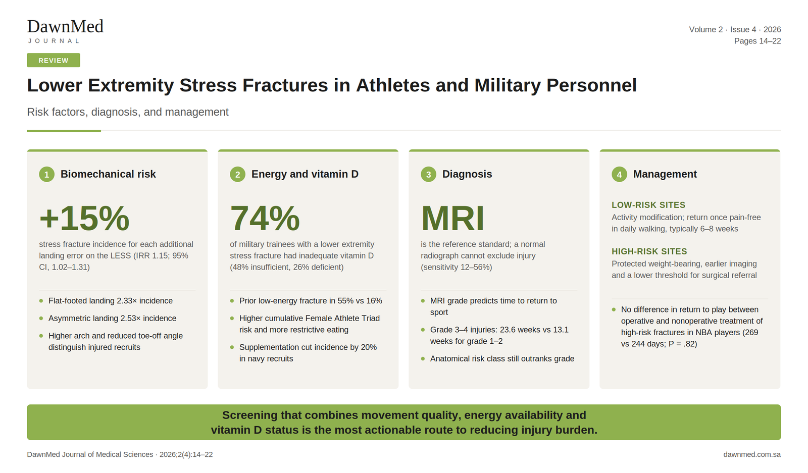
Movement quality has emerged as a measurable and modifiable risk factor. In a prospective cohort of 1,772 military cadets at the United States Military Academy followed for four years, Cameron et al. examined baseline Landing Error Scoring System (LESS) performance against subsequent lower extremity stress fracture [16]. Ninety-four incident stress fractures occurred, a cumulative incidence of 5.3% (95% CI, 4.3%-6.5%). Each additional movement error on the LESS was associated with a 15% increase in the incidence rate of stress fracture (IRR, 1.15; 95% CI, 1.02-1.31; P = .025). Two individual items carried disproportionate weight: participants who consistently landed flat-footed or heel-to-toe had 2.33 times the incidence rate (95% CI, 1.36-3.97; P = .002), and those with asymmetric landing at initial contact had 2.53 times the incidence rate (95% CI, 1.34-4.74; P = .004). All estimates are incidence rate ratios from Poisson models adjusted for sex and cohort entry year.
These findings sit alongside laboratory biomechanics. Pohl et al. reported that female runners with a history of tibial stress fracture exhibited greater peak vertical ground reaction force variables and loading rates than uninjured controls [17]. Milner et al., studying 20 female runners with a history of tibial stress fracture against 20 age- and mileage-matched controls, found significantly greater instantaneous (92.56 ± 24.74 vs 79.65 ± 18.81 BW/s; P = .036) and average (78.97 ± 24.96 vs 66.31 ± 19.52 BW/s; P = .041) vertical loading rates and greater peak tibial shock (7.70 ± 3.21 vs 5.81 ± 1.66 g; P = .014) in the injured group; tibial shock alone correctly classified group membership in 70% of cases, and no difference was found in braking loading rates, tibial varum, or tibial area moment of inertia [18]. Kinematic differences at the hip and rearfoot were described separately by the same group, who reported greater peak hip adduction and greater peak rearfoot eversion in runners with prior tibial stress fracture [19]. The value of the LESS is that it captures related movement deviations without laboratory instrumentation, making it deployable across large groups, and the prospective design of Cameron et al. supports the inference that aberrant movement precedes rather than results from injury.
Foot structure contributes independently. Feng et al. assessed 108 Chinese army recruits before training, deriving the arch index from capacitive plantar pressure footprint imaging and foot kinematics from dynamic gait analysis, then followed them through training; after excluding 10 recruits with other lower limb injuries, 19 who sustained a lower extremity stress fracture were compared with 79 who remained uninjured [20]. Recruits who went on to fracture had a higher arch, reflected in a significantly lower arch index of 0.20 (0.07, 0.24) versus 0.23 (0.17, 0.26); lower values on this index denote a higher arch. They also had a lower toe-off angle (61.59 ± 5.51° versus 64.79 ± 4.79°) and lower landing valgus (eversion) speed, 336.00 (251.02, 428.67) versus 381.20 (313.63, 470.92) °/s. The association was specific to these three variables: landing elevation angle, landing speed, landing varus angle, and valgus amplitude did not differ between groups (all P > .05). Across the whole cohort, arch index correlated positively with landing varus angle (r = 0.25, P < .01) and valgus amplitude (r = 0.14, P < .05). These findings are consistent with Kaufman et al., who identified high arch as a predictor of stress fracture in military personnel [21], and with Williams et al., who found that runners with cavus foot posture experienced higher impact forces and loading rates [22]. Nigg et al. proposed that foot posture modulates impact force transmission, with deviations from neutral alignment concentrating stress at specific skeletal sites [23]. That account explains why a rigid, supinated foot, with little eversion available for shock absorption, transmits more load to the tibia and metatarsals.
3.3.2 Nutritional, metabolic, and hormonal risk factors
Energy availability is the organizing concept in this domain. Gehman et al. studied 51 female runners aged 18 to 36 years, comparing 20 with a history of three or more bone stress injuries against 31 with one or none [24]. The multi-BSI group had significantly higher modified Female Athlete Triad Cumulative Risk Assessment scores (2.90 ± 2.05 versus 1.84 ± 1.59; P = .04), higher Eating Disorder Examination Questionnaire scores (0.92 ± 1.03 versus 0.46 ± 0.49; P = .04), greater body mass fluctuation (15.5% ± 6.5% versus 11.5% ± 4.9%; P = .02), and a markedly greater history of prior low-energy fractures (55% versus 16%; P = .005). The cumulative risk assessment tool used derives from the 2014 Female Athlete Triad Coalition consensus statement, which introduced a risk stratification point system to guide clearance and return-to-play decisions [25].
These observations align with the American College of Sports Medicine position stand, which describes the interrelationships among energy availability, menstrual function, and bone mineral density, with clinical manifestations including disordered eating, functional hypothalamic amenorrhoea, and osteoporosis [7]. Prospective work has since quantified the risk gradient. Barrack et al., in a multisite prospective study of exercising girls and women, found that bone stress injury incidence rose with the number of Triad-related risk factors present, with the strongest risk emerging from combinations rather than any single factor [26]. Tenforde et al. applied the Triad risk stratification prospectively in collegiate athletes and reported that moderate-risk athletes had 2.6 times the risk of bone stress injury (95% CI, 1.3-5.5) and high-risk athletes 3.8 times the risk (95% CI, 1.8-8.0) compared with low-risk athletes, with oligomenorrhoea or amenorrhoea an independent predictor (P = .0069) [27]. The Relative Energy Deficiency in Sport model extends this reasoning beyond female athletes and beyond bone, framing impaired bone health as one of several consequences of sustained low energy availability in athletes of any sex [28]. Subclinical indicators such as body mass fluctuation, restrictive eating attitudes, and menstrual irregularity therefore carry screening value even without a formal eating disorder diagnosis.
Vitamin D status is the second modifiable metabolic factor with consistent evidence. Zalneraitis et al. examined 155 lower extremity stress fractures in military trainees (144 male, 11 female; mean age 22.7 ± 4.85 years) and found that 74% had inadequate 25-hydroxyvitamin D concentrations, 48% insufficient and 26% deficient, with a mean of 26.8 ± 8.37 ng/mL [11]. Fracture location tracked with both vitamin D status and body habitus: patients with foot and ankle fractures had significantly lower 25(OH)D than those with femoral shaft fractures (21.1 versus 30.1 ng/mL; P = .02), while patients with femoral neck and tibial shaft fractures had significantly lower BMI than those with foot and ankle fractures (23.3 versus 27.7, P < .001; and 24.2 versus 27.7, P = .003, respectively). One interpretation is that trabecular-rich distal sites are more sensitive to impaired mineralization [29], although a cross-sectional design cannot establish that sequence.
Supporting military evidence is substantial. Lappe et al. demonstrated in a randomized trial that supplementation with 2,000 mg calcium and 800 IU vitamin D daily through eight weeks of basic training reduced stress fracture incidence by 20% in female navy recruits, independently of other risk factors [30]. Moran et al., following 74 elite male Israeli combat recruits of whom 12 sustained a stress fracture, found that recruits who fractured had significantly lower dietary intake of calcium (589 ± 92 versus 964 ± 373 mg/day; P < .001) and vitamin D (117.9 ± 34.3 versus 157.4 ± 93.3 IU/day; P < .001) at induction [31]. Finestone et al., comparing 36 male and 99 female recruits in a gender-integrated light infantry unit, recorded stress fractures in 12% of the women and none of the men (P = .03) [32]. Training load and biological susceptibility interact here rather than operate independently.
3.4 Diagnosis
Diagnosis begins with clinical suspicion in an athlete or recruit reporting insidious, activity-related, well-localized pain that worsens with loading and eases with rest. Progression of symptoms into daily ambulation, rather than only during sport, signals more advanced injury [33]. Examination typically reveals focal bony tenderness, and pain may be reproduced by direct or indirect percussion, by single-leg hop testing, or by site-specific manoeuvres such as the fulcrum test for femoral shaft injury. Tenderness at the dorsal proximal navicular, the so-called N spot, in an athlete with vague midfoot pain should raise immediate concern for navicular injury, given the diagnostic delays characteristic of that site [34].
Plain radiography remains the usual first investigation but is insensitive early. In a systematic review of imaging accuracy, conventional radiography showed sensitivity ranging from 12% (95% CI, 0-29) to 56% (95% CI, 39-72), with specificity from 88% to 96% [35]. A normal radiograph therefore cannot exclude BSI in a symptomatic athlete, and radiographic changes, when they appear, lag symptom onset by weeks.
Magnetic resonance imaging is the reference standard. The same review reported MRI sensitivity ranging from 68% (95% CI, 45-90) to 99% (95% CI, 95-100), the highest of the modalities assessed, and concluded that MRI is the most sensitive and specific test for lower extremity stress fracture; specificity estimates varied widely, from 4% to 97%, a spread driven by differences in reference standards and populations [35]. MRI has largely displaced nuclear scintigraphy, offering comparable sensitivity with superior anatomic resolution and no ionizing radiation; computed tomography retains a role in characterizing fracture lines and assessing union at sites such as the navicular.
MRI also permits severity grading with prognostic value. Fredericson et al. described a grading system for tibial stress injury based on the progression from periosteal oedema, through marrow oedema visible first on T2-weighted and subsequently on both T1- and T2-weighted sequences, to a discrete fracture line at the most severe grade [33]. Kijowski et al. validated this classification in 142 tibial stress injuries: the most severe grade, characterized by linear intracortical signal abnormality, was associated with significantly longer return to sport, while intermediate grades did not differ from one another (P = .06-.79), supporting an abbreviated classification combining them [36]. Applying this grading approach prospectively in 211 collegiate track and field athletes, Nattiv et al. found that higher MRI grade (P = .004) and lower total-body bone mineral density (P = .030) independently predicted longer recovery: grade 3 and 4 injuries required a mean of 23.6 ± 2.4 weeks to full return to sport versus 13.1 ± 2.0 weeks for grades 1 and 2 (P = .002), and high-grade injuries at trabecular-rich sites such as the femoral neck, sacrum, and pubic bone recovered more slowly still (38.1 ± 6.4 weeks) than those at cortical-rich sites (18.8 ± 2.1 weeks; P = .005) [37]. A subsequent systematic review and meta-analysis provides broader context for grade-informed prognosis [38].
Grading informs but does not replace anatomical risk classification. A low-grade injury at a high-risk site, such as the tension side of the femoral neck or the anterior tibial cortex, warrants more caution than a higher-grade injury at a low-risk site, because the consequence of progression differs fundamentally [5]. Imaging findings should be interpreted jointly with fracture location.
3.5 Management
3.5.1 General principles
Management rests on relative rest, correction of contributing factors, and a graded return to loading. For low-risk injuries, activity modification usually suffices: the athlete stops the provocative activity, maintains fitness through non-impact cross-training, and returns to impact loading once pain-free with daily ambulation, typically over six to eight weeks [15]. Protected weight-bearing in a controlled ankle motion boot or with crutches is used when walking is painful. Return to sport is guided by symptom resolution and progressive load tolerance rather than imaging appearance alone, since marrow oedema can persist beyond clinical recovery.
High-risk injuries follow a different pathway. Non-weight-bearing immobilization, earlier and more liberal advanced imaging, and lower thresholds for surgical referral are appropriate, because delayed union, nonunion, and progression to complete or displaced fracture carry consequences disproportionate to the initial presentation [5]. Concurrent management of the causes is not optional in either pathway: correcting energy availability, addressing vitamin D insufficiency, and modifying training progression are integral to preventing recurrence [39].
3.5.2 Elite athletes
Rizzi et al. described 22 lower extremity stress fractures in 20 NBA players over six seasons [10]. Ten fractures (45%) were treated operatively, with the navicular the most frequently operated site. Players missed a mean of 36.9 ± 35.2 games and 243.0 ± 226.7 days. Among high-risk fractures, there was no significant difference in time to return to play between operative and nonoperative treatment (269.2 versus 243.8 days; P = .82), although the operative group missed substantially more games (59 versus 18.1).
That null result should be read cautiously rather than as evidence of therapeutic equivalence. The comparison is a small, non-randomized subgroup analysis, and allocation was not random: more severe fracture patterns, displaced or chronic injuries, and failed conservative trials plausibly concentrated in the operative group, biasing against surgery. Season timing exerts a further under-appreciated influence, since an injury sustained late in a season may be managed operatively with return deferred to the following season, inflating measured return-to-play time without reflecting biological healing. The intensity of supervision available to professional athletes may also narrow differences that would appear elsewhere.
The wider navicular literature illustrates how unsettled this question remains. Torg et al. first documented that non-weight-bearing cast immobilization succeeded where continued weight-bearing failed [40], and Khan et al. subsequently formalized a protocol of a minimum six weeks of strict non-weight-bearing immobilization followed by graduated rehabilitation, with surgery rarely required when initial treatment is adequate [34]. A meta-analysis by Torg et al. found no statistically significant difference in outcome between non-weight-bearing conservative treatment and surgery (P = .6441), with a non-significant trend favouring conservative management (96% versus 82% successful outcomes), while weight-bearing management was significantly inferior to both [41]. More recent evidence points the other way on some endpoints: in a meta-analysis of 315 navicular stress fractures, Attia et al. reported higher success rates with operative management (97.9% versus 78.1%; OR 5.52; 95% CI, 1.74-17.48; P = .004) and lower refracture rates (1.28% versus 23.53%; P = .047), yet found no significant difference in time to return to play (4.17 versus 4.67 months; P = .60) [42]. Mallee et al., reviewing high-risk stress fractures of the lower leg, found that for navicular fractures surgery was followed by an earlier weighted mean return to sport than conservative treatment (16.4 versus 21.7 weeks), a pattern echoed at the fifth metatarsal base (14 versus 19 weeks). The authors cautioned that the underlying evidence was small and at high risk of bias, and concluded that when navicular fractures are managed conservatively, weight-bearing should be avoided [43].
Taken together, the most consistent advantage of surgery at the navicular lies in union and refracture prevention; its effect on speed of return is unsettled, with Attia et al. finding no significant difference and Mallee et al. an earlier return, a spread compatible with the null return-to-play difference observed by Rizzi et al. What is not in dispute is that continued weight-bearing is inferior to both alternatives.
3.5.3 Pediatric and adolescent patients
Gremillion et al. reviewed 116 lower extremity stress injuries in 97 pediatric and adolescent patients (51 boys, 46 girls; mean age 11.7 years, range 1.1-18) [12]. Nineteen patients (19.6%) sustained high-risk fractures, at the anterior tibia, proximal fifth metatarsal, and femoral neck. These occurred in older children (14.9 versus 11.6 years; P = .01) and required longer return to activity (15 versus 10.5 weeks; P = .027) than low-risk injuries; overall mean return to activity was 11.4 weeks. Radiographs were insufficient for diagnosis in 32% of patients, who required MRI, and that group returned to activity later (16.5 versus 9.5 weeks; P < .01). Younger patients showed a distinct pattern, with a higher incidence of cuboid and calcaneal injuries under 14 years of age. Management was predominantly conservative, most commonly a CAM or walker boot (58.6%) and physical therapy (38.1%), and functional outcomes were favourable, with a mean Lower Extremity Functional Scale score of 73.8.
The age-related distribution has not been extensively characterized in earlier pediatric literature, which has concentrated on adolescent athletes. Risk factor work in this population points toward the same energy-availability and training-load mechanisms seen in adults: Kelsey et al. identified low bone mineral density, menstrual irregularity, and dietary factors as risks among young female cross-country runners [44], and Loud et al. found that higher volumes of high-impact activity correlated with stress fracture in preadolescent and adolescent girls [45]. The frequent use of physical therapy in the Gremillion cohort reflects growing recognition that addressing underlying biomechanical and neuromuscular contributors matters for preventing recurrence, not only for restoring function [39].
3.5.4 Military populations
Military training compresses a large, non-negotiable increase in loading into a fixed period, making recruits a distinctive management population: training cannot always be individualized, and the incentive to conceal symptoms is real. The site distribution reported by Zalneraitis et al., with femoral neck fractures accounting for 60.7% of cases presenting for care, has direct operational implications, since the femoral neck is a high-risk site where missed diagnosis risks displacement and avascular necrosis [11]. A low threshold for MRI in a recruit with groin or thigh pain is justified even when radiographs are normal.
Prevention here has the strongest supporting evidence of any population. Lappe et al. showed that calcium and vitamin D supplementation reduced stress fracture incidence in female navy recruits [30], and dietary inadequacy at induction predicted subsequent fracture among elite combat recruits [31]. Given that 74% of affected trainees in the Zalneraitis cohort had inadequate vitamin D [11], baseline assessment and correction represent a low-cost intervention with a plausible population-level effect. Graded training progression, adequate recovery between loading bouts, and attention to the burden borne by female recruits under standardized regimens [32] complete the preventive picture.
3.6 Prevention and screening
Three screening domains follow from the risk factor evidence, each with a field-deployable instrument.
Movement quality can be assessed with the LESS, which requires only a drop-jump task and video review, scales to large groups, and yields an interpretable dose-response relationship with incidence [16]. Its items point toward specific interventions: flat-footed landing and asymmetric initial contact are trainable through neuromuscular programmes emphasizing landing mechanics and limb symmetry. Whether modifying LESS scores modifies risk has not been established prospectively, which is the principal limitation of using it as more than a stratification tool.
Energy availability and menstrual function should be assessed in female athletes and in male athletes with recognized risk profiles. The Triad Cumulative Risk Assessment offers a structured framework for stratifying risk and informing clearance [25], and the graded relationship between accumulated risk factors and injury incidence supports acting on subclinical findings rather than waiting for diagnostic thresholds [26,27]. Screening questions about weight history, dietary restriction, and menstrual regularity cost nothing, and the REDs framework extends the same logic to male athletes [28].
Vitamin D assessment has the clearest interventional evidence, at least in military populations, where supplementation reduced stress fracture incidence in a randomized trial [30]. Extending that finding to athletes is reasonable but not directly demonstrated, and the association between vitamin D status and fracture site seen in trainees [11] suggests a site-dependent relationship rather than a simple threshold effect [29].
A systematic review with meta-analysis by Wright et al. synthesized risk factors for lower extremity stress fracture in runners and provides context for prioritizing these efforts [46]. Prior BSI deserves particular emphasis among them: it requires no equipment to elicit, and identifies precisely the athletes in whom the other screening domains are most likely to yield actionable findings.
4. Discussion
Read together, the recent studies support a model in which lower extremity BSI emerges from the interaction of mechanical load and biological capacity rather than from any single deficit. On the mechanical side, aberrant landing patterns identified by the LESS [16] and the rigid, high-arched foot described by Feng et al. and earlier work [20-23] each concentrate load on vulnerable sites. On the biological side, low energy availability and its clinical markers [7,24-28] and inadequate vitamin D status [11,29-31] reduce the capacity of bone to absorb that load. The prospective gradient reported by Barrack et al. and Tenforde et al., in which risk rose with the number of accumulated factors rather than with any one of them [26,27], is the clearest quantitative expression of this interaction.
The management evidence turns on anatomical risk classification more than on any single treatment comparison [5,15]. At the navicular, the apparent conflict between meta-analyses narrows once endpoints are separated: operative fixation shows a consistent advantage in union and refracture prevention [42], while its effect on speed of return is inconsistent across syntheses [41-43] and absent in the professional basketball cohort [10]. The one conclusion shared by every source is that continued weight-bearing during conservative treatment of navicular fractures is inferior [40,41,43].
For practice, the findings assemble into a screening pathway that requires little equipment: a field movement screen [16], structured assessment of energy availability and menstrual function [25-27], and measurement of 25-hydroxyvitamin D with correction of insufficiency [11,30]. Prior bone stress injury, elicited by history alone, identifies the athletes in whom these screens are most likely to change management [46].
The gaps are equally clear. No randomized comparison of operative and nonoperative management exists for any high-risk site, and the available return-to-play comparisons are confounded by indication [10]. Whether correcting a modifiable screen result lowers subsequent injury risk has direct randomized support only for calcium and vitamin D supplementation in military recruits [30]; for movement retraining and energy availability intervention, that step remains to be demonstrated [16]. Prospective multicentre cohorts that integrate biomechanical and biological measurement, and trials that intervene on screen-positive athletes, are the studies this field most needs.
4.1 Limitations
This is a narrative review, and its selection of literature was not systematic. Articles were chosen for relevance rather than through a predefined, reproducible protocol, which introduces potential selection bias and means relevant work may have been omitted. No formal quality appraisal was performed, so the evidence summarized here is not weighted by methodological quality; the recent evidence base is small, observational, and in several instances drawn from single centres. Populations differ substantially across the studies discussed, spanning military recruits, professional basketball players, female distance runners, and pediatric patients, and findings from one do not transfer straightforwardly to another. Several key statistics derive from cross-sectional or retrospective designs that cannot establish temporal sequence, and no randomized comparison of operative versus nonoperative management of high-risk stress fractures exists.
5. Conclusion
Lower extremity bone stress injuries arise from the interaction of biomechanical, nutritional, metabolic, and demographic factors, and no single factor explains them. The division of anatomical sites into low-risk and high-risk categories remains the most consequential decision in management, determining diagnostic urgency, weight-bearing status, and the threshold for surgical referral. Outcomes are generally favourable with conservative care at low-risk sites and in pediatric patients, while at high-risk sites recent navicular evidence suggests surgery improves union and reduces refracture without a consistent effect on time to return. Screening that combines movement quality, energy availability, and vitamin D status, coupled with targeted correction of what it finds, is the most immediately actionable route to reducing injury burden in athletic and military populations.
Table 1. Summary of key recent studies (2022–2023)
| Study / Country | Design | Population and sample size | Key findings |
|---|
| Feng et al., 2022 [20] / China | Case-control (pre-training assessment, prospective follow-up) | 108 army recruits assessed before training; after excluding 10 with other lower limb injuries, 19 who sustained a lower extremity stress fracture were compared with 79 uninjured controls. Age, sex and BMI not reported in the source. | Fracture group had a higher arch, reflected in a significantly lower arch index (lower values denote a higher arch; 0.20 [0.07, 0.24] vs 0.23 [0.17, 0.26]); lower toe-off angle (61.59 ± 5.51° vs 64.79 ± 4.79°); lower landing valgus speed (336.00 [251.02, 428.67] vs 381.20 [313.63, 470.92] °/s). Differences reported collectively as P < .05 or .01. Landing elevation angle, landing speed, landing varus angle and valgus amplitude did not differ (all P > .05). Across the cohort, arch index correlated with landing varus angle (r = 0.25, P < .01) and valgus amplitude (r = 0.14, P < .05). |
| Cameron et al., 2022 [16] / USA | Prospective cohort, 4-year follow-up | 1,772 incoming military academy cadets. Mean age 18.7 ± 0.9 years (range, 17-23); 1,178 men (66%), 594 women (34%); mean BMI 23.9 ± 2.8 kg/m². | 94 incident lower extremity stress fractures; cumulative incidence 5.3% (95% CI, 4.3%-6.5%). Each additional LESS movement error: IRR 1.15 (95% CI, 1.02-1.31; P = .025). Flat-footed landing: 2.33 (95% CI, 1.36-3.97; P = .002). Asymmetric landing at initial contact: 2.53 (95% CI, 1.34-4.74; P = .004). |
| Gehman et al., 2022 [24] / USA | Cross-sectional | 51 female runners aged 18-36 years (mean age 26.2 ± 4.2 years; mean BMI 21.6 ± 2.0 kg/m²): 20 with a history of ≥3 bone stress injuries (multiBSI) vs 31 with ≤1 (controls). | Multi-BSI group had higher modified Triad CRA scores (2.90 ± 2.05 vs 1.84 ± 1.59; P = .04), higher EDE-Q scores (0.92 ± 1.03 vs 0.46 ± 0.49; P = .04), greater body mass fluctuation (15.5% ± 6.5% vs 11.5% ± 4.9%; P = .02), and more prior low-energy fractures (55% vs 16%; P = .005). |
| Rizzi et al., 2022 [10] / USA | Case series, 6 NBA seasons (2013-2014 to 2018-2019) | 20 professional basketball players with 22 lower extremity stress fractures. Mean age 25.4 ± 4.8 years; all male; mean BMI 25.1 ± 1.5 kg/m². | Incidence 0.12 stress fractures per 1000 player-games. Foot 17/22 (77%); navicular the most common single site (8, 36%); tibia 5 (23%). Operative treatment in 45% (10/22). Mean 36.9 ± 35.2 games and 243.0 ± 226.7 days missed. Among high-risk fractures, no significant difference in return to play between operative and nonoperative treatment (269.2 vs 243.8 days; P = .82). |
| Zalneraitis et al., 2023 [11] / USA | Descriptive cross-sectional | 155 lower extremity stress fractures in military trainees. Mean age 22.7 ± 4.85 years (range, 17-42); 144 men (92.9%), 11 women (7.1%); BMI 23.3-27.7 kg/m² across fracture sites. | 74% had inadequate 25(OH)D (48% insufficient, 26% deficient); mean 26.8 ± 8.37 ng/mL. Sites: femoral neck 94 (60.7%), tibial shaft 34 (21.9%), foot/ankle 14 (9.0%), femoral shaft 13 (8.4%). Foot/ankle vs femoral shaft 25(OH)D 21.1 vs 30.1 ng/mL (P = .02). Lower BMI in femoral neck (23.3) and tibial shaft (24.2) than foot/ankle fractures (27.7); P < .001 and P = .003. |
| Gremillion et al., 2023 [12] / USA | Retrospective case series | 97 pediatric and adolescent patients (<18 years) with 116 stress injuries at a tertiary children's hospital. Mean age 11.7 years (range, 1.1-18); 51 boys (52.6%), 46 girls (47.4%); mean BMI 21.9 kg/m² (range, 13.4-44.0). | Tibia most common (33 of 116 injuries, 28.4%), cuneiforms least (4, 3.4%); high-risk fractures in 19 (19.6%). Patients <14 years more likely to sustain cuboid and calcaneal injuries. High-risk fractures occurred in older children (14.9 vs 11.6 years; P = .01) and required longer return to activity (15 vs 10.5 weeks; P = .027); overall mean 11.4 weeks. CAM/walker boot 58.6%, physical therapy 38.1%. Mean LEFS 73.8. |
BSI, bone stress injury; CRA, Cumulative Risk Assessment; EDE-Q, Eating Disorder Examination Questionnaire; IRR, incidence rate ratio; LEFS, Lower Extremity Functional Scale; LESS, Landing Error Scoring System; NBA, National Basketball Association; 25(OH)D, 25-hydroxyvitamin D. Values are mean ± SD or median (interquartile range) as reported in the source publication.
Declarations
Ethics approval and consent to participate. Not applicable. This review synthesizes previously published literature and involved no new data collection from human participants or animals.
Consent for publication. Not applicable.
Availability of data and materials. All data discussed in this review are available in the cited published sources.
Competing interests. The authors declare that they have no competing interests.
Funding. This research received no specific grant from any funding agency in the public, commercial, or not-for-profit sectors.
Authors' contributions. All authors contributed to the conception and design of the review. The literature search and data extraction were performed by all authors. The first draft of the manuscript was written by all authors, who commented on subsequent versions. All authors read and approved the final manuscript.
Acknowledgements. None.