Jomana Nasser Alkhraisi¹, Waad Mohammed Abu Nakha², Deyala Riyadh Alzaheb¹, Sarah Alomran³, Ghala Tawfiq Aldarwish³, Joud Abdulhafith A. AlAhmady⁴, Waleed Badoghaish¹*.Lemmel Syndrome Masquerading as an Ampullary Mass: A Case Report and Mini-Review. DawnMed Journal of Medical ScienceVolume 2, Issue 4:p: 31-37,2026.
1 Faculty of Medicine, University of Tabuk, Tabuk, Saudi Arabia.
2 College of Medicine, King Saud University, Riyadh, Saudi Arabia.
3 College of Medicine, King Saud bin Abdulaziz University for Health Sciences, Riyadh, Saudi Arabia.
4 College of Medicine, Taibah University, Medina, Saudi Arabia.
Corresponding author
*Waleed Badoghaish
Faculty of Medicine, University of Tabuk, Tabuk, Saudi Arabia.
Email: Wbadoghaish@gmail.com
Received: July 21, 2026 | Peer reviewed: September 3, 2026 | Accepted: September 18, 2026 | Published: October 9, 2026
Cite this article as
Alkhraisi JN, Abu Nakha WM, Alzaheb DR, Alomran S, Aldarwish GT, AlAhmady JAA, et al. Lemmel Syndrome Masquerading as an Ampullary Mass: A Case Report and Mini-Review. DawnMed Journal 2(4):31–37 https://doi.org/10.64039/djms.2026.____
Background: Lemmel syndrome is an uncommon cause of obstructive jaundice due to a periampullary duodenal diverticulum. Its diagnosis can be challenging when the diverticulum mimics an ampullary mass.
Case Presentation:
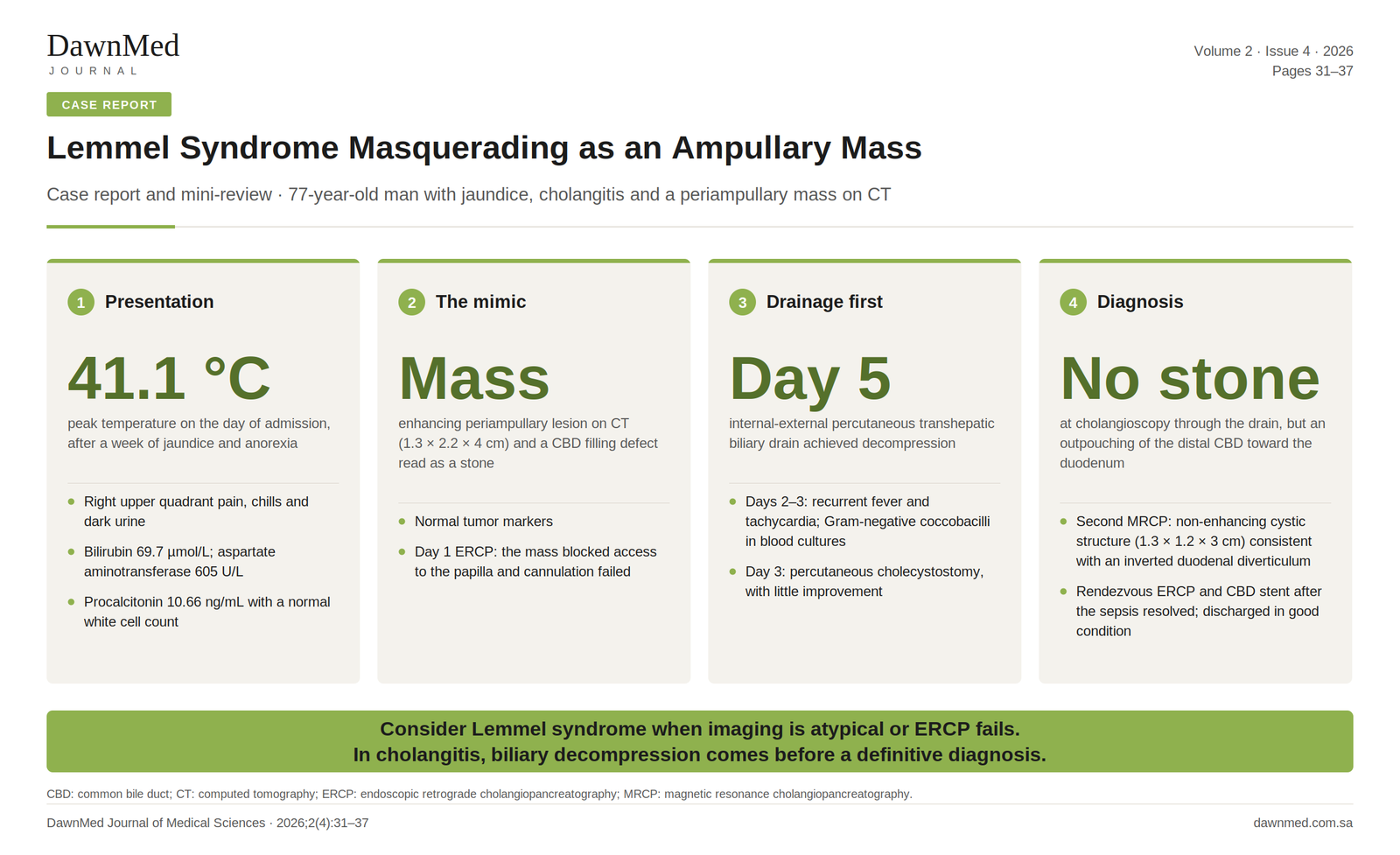
A 77-year-old man presented with jaundice, anorexia, fever, chills, and right upper quadrant pain. Laboratory investigations showed a mixed cholestatic and hepatocellular pattern of liver injury with elevated inflammatory markers. Computed tomography (CT) demonstrated an enhancing polypoid lesion involving the ampulla, distal common bile duct dilatation with a filling defect read as a stone, mild pancreatic duct dilatation, and multiple large gallbladder stones, raising concern for an ampullary tumor and choledocholithiasis. Initial endoscopic retrograde cholangiopancreatography (ERCP) failed because the suspected mass obstructed access to the papilla. With recurrent fever, tachycardia, blood cultures positive for Gram-negative coccobacilli, and worsening inflammatory and cholestatic markers, percutaneous transhepatic biliary drainage was performed after limited improvement with cholecystostomy. A subsequent cholangiogram and cholangioscopy demonstrated a distal common bile duct outpouching without a stone. A dedicated repeat magnetic resonance cholangiopancreatography (MRCP) identified a non-enhancing, non-restricting periampullary cystic structure consistent with an inverted duodenal diverticulum, supporting the diagnosis of Lemmel syndrome. A subsequent rendezvous ERCP was successfully performed with common bile duct stent placement, and he was discharged in good condition.
Conclusion:
This case shows the diagnostic challenge of Lemmel syndrome when a periampullary diverticulum mimics malignancy on initial imaging, and the role of repeat dedicated imaging, cholangioscopy, percutaneous biliary drainage, and rendezvous ERCP when conventional ERCP is unsuccessful.
Introduction
Lemmel syndrome is obstructive jaundice caused by extrinsic compression of the distal common bile duct (CBD) by a periampullary duodenal diverticulum, in a patient without choledocholithiasis or a pancreaticobiliary tumor [1,2]. Periampullary diverticula are common. Endoscopic series report them in 7.5% to 32.8% of patients undergoing endoscopic retrograde cholangiopancreatography (ERCP), and patients with a diverticulum are older than those without [3,4]. Most are asymptomatic and are found incidentally [3], and biliary obstruction from the diverticulum itself is an unusual complication [2].
The presentation is non-specific and overlaps with choledocholithiasis and malignant biliary obstruction [2,5]. Computed tomography (CT) and magnetic resonance cholangiopancreatography (MRCP) are the main non-invasive tests, and ERCP is used for diagnosis and treatment when the papilla can be reached [6]. Even so, a diverticulum filled with fluid or stones can look like a soft-tissue mass. Maloku and Aybay reported two patients whose diverticulum was read as a pancreatic head tumor, and in one of them MRCP revealed the diagnosis only after three failed stent changes [7]. In another report, a 6-cm duodenal mass that could not be reached endoscopically proved at surgery to be a stone-filled diverticulum [8].
There is no standardized approach to treatment [1]. Most patients improve with conservative or endoscopic treatment [6], and surgery is reserved for endoscopic failure or complications [5].
We report a 77-year-old man whose periampullary diverticulum was read as an ampullary mass on CT. The first ERCP could not reach the papilla, cholangitis needed percutaneous drainage, and the diagnosis followed cholangioscopy and a second, dedicated MRCP. We also review recently reported cases.
Case Presentation
History
A 77-year-old man presented to the emergency department in July 2025 with a one-week history of anorexia and jaundice. His medical history included type 2 diabetes mellitus (sitagliptin 100 mg once daily), hypertension (lisinopril 5 mg once daily), dyslipidemia (atorvastatin 10 mg once daily), and benign prostatic hyperplasia (tamsulosin 0.4 mg once daily). He had no known drug allergies and no family history of malignancy or hepatitis.
His family first noticed yellow discoloration of his eyes and a falling appetite about a week before admission, and his oral intake continued to decline. The day before presentation he had two episodes of non-bloody, non-bilious vomiting containing food particles. On the day of admission he developed subjective fever, chills, right upper quadrant pain, reduced activity, dark urine, nausea, and diaphoresis. He reported no night sweats or weight loss, and the systemic review was otherwise negative for cardiovascular, respiratory, gastrointestinal, urinary, neurological, and skin symptoms.
Examination
He looked unwell and was jaundiced, but was alert, oriented, and not in respiratory distress. Heart sounds were normal with a regular pulse, the chest was clear, and there was no focal neurological deficit, lymphadenopathy, or leg edema. The abdomen was soft with right upper quadrant tenderness, Murphy’s sign was negative, and there were no stigmata of chronic liver disease.
On arrival (15:31) his temperature was 39.4°C, heart rate 137 beats/min, blood pressure 151/44 mmHg, and oxygen saturation 94%. At 16:00 his temperature reached 41.1°C, with a heart rate of 128 beats/min and an oxygen saturation of 92%. His last readings in the emergency department, at 22:13, were a temperature of 36.6°C, heart rate 96 beats/min, blood pressure 131/52 mmHg, and oxygen saturation 97%.
Investigations
Admission blood tests (Table 1) showed a mixed cholestatic and hepatocellular pattern, with total bilirubin 69.7 µmol/L, aspartate aminotransferase (AST) 605 U/L, and alanine aminotransferase (ALT) 309 U/L. C-reactive protein (CRP) and procalcitonin were high, while the white cell count was normal with lymphopenia. Venous lactate was 2.81 mmol/L and was 1.13 mmol/L when repeated at 21:02. Creatinine was 90 µmol/L, with an estimated glomerular filtration rate of 75 mL/min/1.73 m². Random glucose was 24.7 mmol/L, and urinalysis showed glucosuria, ketonuria, and microscopic hematuria without leukocyte esterase or nitrites; venous pH was 7.42. Troponin I was below the assay threshold on two samples, and B-type natriuretic peptide and serum amylase were normal. Alpha-fetoprotein (AFP), carcinoembryonic antigen (CEA), and carbohydrate antigen 19-9 (CA 19-9) were within normal limits.
Contrast-enhanced CT of the abdomen and pelvis on the day of admission reported an enhancing polypoid soft-tissue mass projecting from the second part of the duodenum and reaching the third part. It measured 1.3 × 2.2 × 4 cm and involved the ampulla. The CBD was dilated to 8 mm with an intraluminal filling defect reported as a stone, the intrahepatic ducts were mildly dilated, and the main pancreatic duct measured 5 mm. The gallbladder was distended and contained multiple large stones, without signs of acute cholecystitis. The pancreas was otherwise unremarkable, and there was no lymphadenopathy, free fluid, or metastatic disease. Incidental findings were bilateral renal cortical scarring, an enlarged prostate with a trabeculated bladder, and a small sliding hiatal hernia.
The differential diagnosis at this stage is summarized in Table 2.
Table 2. Differential diagnosis at presentation
Diagnosis
For
Against
Choledocholithiasis
Jaundice, dark urine, fever, RUQ pain; raised GGT and ALP; gallbladder stones; CBD filling defect on CT
A stone did not explain the enhancing periampullary soft-tissue lesion
Ampullary tumor
Progressive jaundice, anorexia, older age; enhancing periampullary mass on CT
Normal CA 19-9, CEA, and AFP; no lymphadenopathy or metastatic disease on CT
Pancreatic head tumor
Jaundice, anorexia, older age; CBD and pancreatic duct dilatation
No pancreatic head enlargement or mass on CT; normal CA 19-9
Acute hepatitis
Jaundice, fever, RUQ pain, nausea and vomiting; raised ALT and AST
No sick contacts; dilated bile ducts on CT
RUQ: right upper quadrant; CBD: common bile duct; GGT: gamma-glutamyl transferase; ALP: alkaline phosphatase; ALT: alanine aminotransferase; AST: aspartate aminotransferase; CT: computed tomography; CA 19-9: carbohydrate antigen 19-9; CEA: carcinoembryonic antigen; AFP: alpha-fetoprotein.
Hospital course
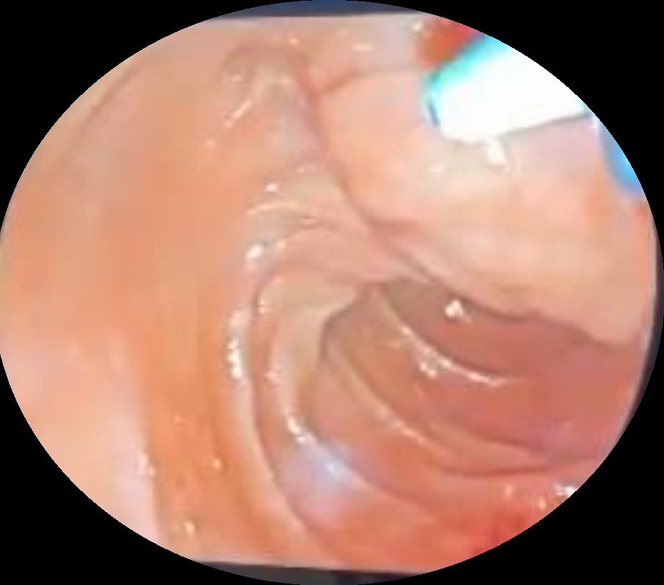
Intravenous meropenem was started on the day of presentation (Table 3). On day 1, after clinical and biochemical improvement, ERCP was attempted, but a mass obstructed access to the papilla and cannulation failed (Figure 1). A repeat attempt with stent placement was planned.
Figure 1. Endoscopic view of the periampullary region during the first ERCP attempt (day 1).
On days 2 and 3, despite meropenem, he developed recurrent high-grade fever, tachycardia, and repeated vomiting, with rising inflammatory markers, gamma-glutamyl transferase (GGT), and alkaline phosphatase (ALP). Blood cultures grew Gram-negative coccobacilli. On the advice of the infectious diseases team, doxycycline and gentamicin were added to meropenem, and Brucella serology and a blood-culture identification panel were sent. The repeat ERCP was cancelled because of his instability. A percutaneous cholecystostomy was placed on day 3 but gave little improvement, and on day 5 interventional radiology placed an internal-external percutaneous transhepatic biliary drain, which achieved decompression.
On day 12 an MRCP was attempted, but the patient did not complete the study, and only T2-weighted and diffusion-weighted sequences were obtained. The periampullary structure could not be characterized, although there was no obvious diffusion restriction at the site of the suspected lesion. The report described a small subcapsular hepatic hematoma, noted as previously seen, and a loculated right pleural collection with restricted diffusion that could represent a hemothorax. Neither had been described on the admission CT.
Upper gastrointestinal surgery reviewed the patient and referred him to hepatobiliary surgery, whose impression was obstructive jaundice from an ampullary mass. They advised upper endoscopy with biopsy of the suspected mass once the sepsis had settled, and removal of the presumed CBD stone through the existing drain tract. Interventional radiology exchanged the drain over a guidewire and passed an 11 French (Fr) sheath into the CBD. The cholangiogram showed a filling defect in the distal CBD. SpyGlass cholangioscopy through the sheath showed no stone; instead there was an outpouching of the distal CBD toward the duodenum, confirmed by contrast injection. The sheath was removed and a new 10 Fr biliary drainage catheter was placed, and the patient tolerated the procedure without immediate complications.
Endoscopic treatment was still not possible, so upper gastrointestinal surgery was consulted again and requested a repeat MRCP, because the first study had been limited by poor cooperation. This was performed after a follow-up CT on day 23, using contrast-enhanced liver-mass and MRCP protocols. It showed a periampullary cystic structure measuring 1.3 × 1.2 × 3 cm without post-contrast enhancement or restricted diffusion, consistent with an inverted duodenal diverticulum. There was no focal suspicious lesion, lymphadenopathy, obstructing CBD stone, or intrahepatic duct dilatation, and the radiological impression was obstructive periampullary inverted duodenal diverticulum (Lemmel syndrome). The subcapsular hepatic collection had decreased to 1 × 2 cm, and the right pleural collections persisted and were considered likely to represent hemothorax. The MRCP studies also reported hepatic and splenic iron overload, a right renal angiomyolipoma, bilateral renal cortical scarring, and heterogeneous signal in the spine at several levels.
After the sepsis had resolved, a rendezvous ERCP was performed and a CBD stent was placed. He recovered clinically and his biochemical markers normalized. He was discharged in good condition with the CBD stent and cholecystostomy tube in place, and clinic review was planned within four weeks for their removal and a decision on definitive surgery.
Table 3. Timeline of the admission
Day (date)
Findings and interventions
Day 0 (19 July 2025)
Presentation with jaundice, fever, and right upper quadrant pain. CT: enhancing periampullary mass, CBD filling defect read as a stone, gallbladder stones. Intravenous meropenem started.
Day 1 (20 July 2025)
First ERCP: mass obstructing access to the papilla; cannulation failed.
Days 2–3 (21–22 July 2025)
Recurrent fever, tachycardia, and vomiting; rising CRP, GGT, and ALP. Blood cultures: Gram-negative coccobacilli. Doxycycline and gentamicin added; Brucella serology and blood-culture identification panel sent. Repeat ERCP cancelled.
Day 3 (22 July 2025)
Percutaneous cholecystostomy, with little clinical improvement.
First MRCP, incomplete. Small subcapsular hepatic hematoma (noted as previously seen) and loculated right pleural collection (possible hemothorax).
After day 12
Surgical review. Cholangiogram through the drain: distal CBD filling defect. SpyGlass cholangioscopy: no stone; distal CBD outpouching toward the duodenum. Drain exchanged for a 10 Fr catheter.
Day 23 (11 August 2025)
Follow-up CT of the abdomen and pelvis.
Day 23 or later
Second MRCP: non-enhancing, non-restricting periampullary cystic structure (1.3 × 1.2 × 3 cm) consistent with an inverted duodenal diverticulum; hepatic collection 1 × 2 cm; right pleural collections persisting.
Before discharge
Sepsis resolved. Rendezvous ERCP with CBD stent. Discharged with CBD stent and cholecystostomy tube; clinic review planned within 4 weeks.
CT: computed tomography; CBD: common bile duct; ERCP: endoscopic retrograde cholangiopancreatography; CRP: C-reactive protein; GGT: gamma-glutamyl transferase; ALP: alkaline phosphatase; MRCP: magnetic resonance cholangiopancreatography.
Discussion and Mini-Review
Definition and mechanism
The syndrome was first described in 1934 [9]. Periampullary diverticula are mucosal outpouchings without a muscular layer, lying within 2 to 3 cm of the ampulla of Vater [10]. Three mechanisms of obstruction have been proposed: compression of the distal CBD or ampulla by the diverticulum, fibrosis and scarring of the ampulla after diverticulitis, and dysfunction of the sphincter of Oddi [10]. Stones and tumor must be excluded before the diagnosis is made [1,2], and gallstones, bile duct stones, and cholangitis are themselves more common in patients with duodenal diverticula [11].
Papilla position and cannulation
Boix and colleagues classified periampullary diverticula by the position of the major papilla: type I, inside the diverticulum; type II, at its margin; and type III, near it [4]. In their series a diverticulum did not make deep cannulation more difficult [4]. In a prospective series of 601 patients, cannulation succeeded in 94.9% of those with a diverticulum and 94.8% of those without once the papilla had been found, but the papilla could not be found in 8.3% of patients with a diverticulum compared with 0.9% of those without [12]. A 2024 meta-analysis of 28 studies found that an intradiverticular papilla increased the rate of difficult cannulation (odds ratio 1.60, 95% confidence interval 1.05–2.44), whereas diverticula overall were no longer associated with cannulation failure in studies published after 2015 [13]. Our patient’s papilla could not be reached at the first ERCP.
Clinical presentation
Symptoms are non-specific. In a pooled review of 46 cases, abdominal pain was reported in 80%, jaundice in 54%, fever in 50%, nausea or vomiting in 32%, and weight loss in 15% [9]. In an earlier review of 17 cases, abdominal pain was present in 64.7% and fever in 52.9% [1]. All 12 patients in a single-center cohort had clinical or biochemical evidence of obstructive jaundice [6]. Our patient had pain, jaundice, and fever, with a normal white cell count at presentation and Gram-negative coccobacilli in the blood cultures.
Reported cases
Table 4 lists twelve selected reports published from 2022 to 2026 beside our case. A diverticulum was first taken for a tumor or mass in three patients from two of these reports: as a pancreatic head tumor in both patients of Maloku and Aybay [7], and as a 6-cm duodenal mass in the patient of Wautelet et al. [8]. As in our patient, the diagnosis in the second patient of Maloku and Aybay came only after repeated procedures. Vikash et al. and Krisem et al. each described a double-duct sign, with simultaneous dilatation of the CBD and pancreatic duct [2,14]. The CBD (8 mm) and main pancreatic duct (5 mm) were also both dilated in our patient. Buhusayen et al. reported cholangitis with bacteremia and gallstones and considered Lemmel syndrome the most likely diagnosis [15], and Battah et al. described cholangitis without biliary dilatation [16].
Table 4. Selected reported cases of Lemmel syndrome and the present case
Report
Age/sex
Presentation
Distinguishing feature
Love et al., 2022 [1]
82 F
Acute abdominal pain and vomiting
Presented as gastric outlet obstruction; liver tests normal
Shrivastava et al., 2023 [11]
80 M
Upper abdominal discomfort; obstructive jaundice
Papilla inside the diverticulum; abrupt termination of the CBD and pancreatic duct at the thickened diverticular wall on CT
Battah et al., 2023 [16]
58 M
Abdominal pain, nausea, and vomiting; cholangitis
Papilla entirely inside a large diverticulum; cholangitis without biliary dilatation; precut sphincterotomy and stent
Krisem et al., 2023 [2]
82 M
Intermittent abdominal pain (2 months) and jaundice
Large air-filled diverticulum (8.3 cm) compressing the distal CBD and pancreatic duct; double-duct sign
Maloku and Aybay, 2023 [7]
82 M; 48 (NR)
Abdominal pain, vomiting, fever, and jaundice
Both read as pancreatic tumors; the second patient had papillotomy, stenting, and three failed stent changes before MRCP showed the diverticulum
Sergi et al., 2024 [17]
79 F
Cholangitis and obstructive jaundice
Two diverticula, papilla near the larger (type III); ERCP sphincterotomy failed and a laparoscopic rendezvous was performed
Vikash et al., 2024 [14]
76 M
Epigastric pain; cholestatic liver tests
Double-duct sign; one medium-sized diverticulum at upper endoscopy
Ali et al., 2024 [10]
60 F
Abdominal discomfort and vomiting
Conservative treatment for diverticulitis, then bypass surgery with cholecystectomy
Wautelet et al., 2024 [8]
67 (NR)
6-cm duodenal mass with biliary stenosis; previous gastric bypass
Endoscopic access impossible; a stone-filled diverticulum was found at surgery
Buhusayen et al., 2025 [15]
74 M
Abdominal pain and fever; abnormal liver tests and leukocytosis
Bacteremia and multiple gallstones without biliary dilatation; Lemmel syndrome considered the most likely diagnosis
Hanaoka et al., 2025 [18]
67 M
Recurrent liver dysfunction; hepatocellular injury with cholestasis
CT: diverticulum compressing the distal bile duct, no stone; sludge-like bile after sphincterotomy and balloon sweeping; liver tests improved quickly
Ben Ismail et al., 2026 [5]
71 M
Right upper quadrant pain, progressive jaundice, and weight loss for 2 months
CT showed a diverticulum compressing the distal CBD; ERCP with sphincterotomy and stent led to improvement
Present case
77 M
Jaundice, cholangitis, and a periampullary mass on CT
First ERCP failed; diagnosis after percutaneous drainage, cholangioscopy, and a second MRCP; rendezvous ERCP
F: female; M: male; CBD: common bile duct; CT: computed tomography; MRCP: magnetic resonance cholangiopancreatography; ERCP: endoscopic retrograde cholangiopancreatography; NR: not reported.
Imaging and the mass-like appearance
Ultrasound can show biliary dilatation [19] but frequently fails to establish the diagnosis [6]. CT is regarded as the reference imaging test for a diverticulum compressing the intrapancreatic CBD [19], and CT and especially MRCP show the relationship between the diverticulum and the biliary tree [6]. The diverticulum may appear as an outpouching from the medial wall of the second part of the duodenum, filled with air or contrast [2,11]. One filled with fluid or stones can be mistaken for a mass [7,8]. In our patient CT showed an enhancing polypoid lesion, whereas the second MRCP showed a cystic structure without enhancement or restricted diffusion, which argued against a solid tumor.
The CT and MRCP measurements differed (1.3 × 2.2 × 4 cm and 1.3 × 1.2 × 3 cm), and we take them to describe the same periampullary structure. The studies were at least 23 days apart, biliary drainage and treatment of sepsis took place in between, and CT measured an enhancing lesion where MRCP measured a cystic one. The CT report described the lesion as projecting from the second part of the duodenum and reaching the third part, a long extension for a diverticulum defined as lying within 2 to 3 cm of the ampulla. A diverticulum inverted into the duodenal lumen can extend distally along it, which may explain this.
Passed common bile duct stone
A passed CBD stone is the main alternative diagnosis. The gallbladder contained multiple large stones, the admission CT reported a stone in the distal CBD, and he presented with cholangitis. In choledocholithiasis, aminotransferases can exceed 1,000 U/L and fall within 3 to 14 days after treatment [20], so an AST of 605 U/L and an ALT of 309 U/L do not separate a stone from a diverticulum. Hanaoka et al. also described recurrent hepatocellular injury attributed to diverticular compression without a stone [18].
Against a passed stone, GGT and ALP kept rising on days 2 and 3, which points to continuing obstruction, and there was little improvement with antibiotics or cholecystostomy before percutaneous transhepatic drainage. The filling defect was still present in the distal CBD at cholangiography through the drain. Cholangioscopy at the same procedure found no stone but showed an outpouching of the distal CBD toward the duodenum, and the second MRCP showed no CBD stone. All of these studies followed biliary decompression, however, so a small stone that passed earlier cannot be excluded. We regard Lemmel syndrome as the most likely diagnosis, not a proven one.
Management
ERCP with sphincterotomy is the commonest treatment for Lemmel syndrome, and interventional radiology or surgery is considered when endoscopic management fails [17]. After a failed ERCP, percutaneous transhepatic drainage can be followed by a percutaneous-endoscopic rendezvous procedure [21], and a laparoscopic rendezvous has also been used [17]. In a small series of patients with retained bile duct stones, Odabasi et al. proposed that an antegrade guidewire passed through a T-tube and retrieved through the duodenoscope would increase ERCP success, especially with large diverticula or an intradiverticular papilla [22]. Surgical options include excision of the diverticulum [11] and bypass [10].
In our patient, with sepsis and a failed first ERCP, drainage took priority over a definitive diagnosis. After percutaneous transhepatic drainage and resolution of the sepsis, a rendezvous ERCP allowed internal stenting, and no surgery was needed during the admission. Whether he needs definitive surgery will be decided at follow-up.
Procedure-related complications
A subcapsular hepatic hematoma and a loculated right pleural collection were reported on day 12, after the cholecystostomy on day 3 and the transhepatic drain on day 5, and neither had been described on the admission CT. Subcapsular hematomas after percutaneous biliary procedures are usually self-limiting and often lie at the catheter entry site, and a transhepatic approach through the intercostal space can injure an intercostal artery and present as hemothorax [23]. In a prospective CT study of 31 percutaneous biliary procedures, abnormalities were found after 14 procedures (45%), including subcapsular hematoma and subcapsular or perihepatic fluid, and only five were associated with clinically apparent complications [24]. Both collections may therefore have been related to the procedures, although we cannot confirm this. The hepatic collection had decreased on the second MRCP, and the pleural collection persisted.
Limitations
This report describes one patient, and a passed stone cannot be excluded entirely. The review is not systematic, so Table 4 does not include all published cases.
Conclusion
Lemmel syndrome is an uncommon cause of obstructive jaundice that should be considered when imaging findings are atypical or ERCP fails, because it can closely mimic choledocholithiasis or periampullary malignancy. A periampullary diverticulum may appear as an ampullary mass on cross-sectional imaging, and diagnosis and treatment may need a staged approach with dedicated MRCP, cholangioscopy, and rendezvous ERCP. In patients with cholangitis or sepsis, biliary decompression should take priority over a definitive diagnosis, and percutaneous transhepatic drainage can be an effective rescue option when ERCP fails or is not feasible. A high index of suspicion and an individualized multidisciplinary approach help avoid misdiagnosis and delay.
Declarations
Consent for Publication: Written informed consent was obtained from the patient for publication of this case report and the accompanying image.
Author Contributions: All authors contributed to data collection, literature review, drafting, and critical revision of the manuscript, and approved the final version.
Data Availability: All data relevant to this case are included in the article.
Acknowledgements: We thank the gastroenterology, interventional radiology, hepatobiliary surgery, upper gastrointestinal surgery, and infectious diseases teams who cared for the patient.
Conflicts of Interest: None declared.
Funding: None.
Use of Artificial Intelligence: The authors used AI only for language editing and reviewed and checked all content.
References
Love JS, Yellen M, Melitas C, Yazici C, Zar F. Diagnosis and management of Lemmel syndrome: an unusual presentation and literature review. Case Rep Gastroenterol. 2022;16(3):663–674. doi:10.1159/000528031. PMID: 36605730.
Krisem M, Hirunpat P, Tungtrongchitr N. Lemmel syndrome, a rare cause of obstructive jaundice by periampullary duodenal diverticulum: case report and review of the literature. J Clin Imaging Sci. 2023;13:11. doi:10.25259/JCIS_9_2023. PMID: 37152440.
Boix J, Lorenzo-Zúñiga V, Añaños F, Domènech E, Morillas RM, Gassull MA. Impact of periampullary duodenal diverticula at endoscopic retrograde cholangiopancreatography: a proposed classification of periampullary duodenal diverticula. Surg Laparosc Endosc Percutan Tech. 2006;16(4):208–211. doi:10.1097/00129689-200608000-00002. PMID: 16921297.
Ben Ismail I, Manai G, Torkhani A, Sghaier M, Zenaidi H, Zoghlami A. Periampullary duodenal diverticulum causing obstructive jaundice: a case of Lemmel syndrome and review of the literature. Int J Surg Case Rep. 2026;138(3):448–452. doi:10.1097/RC9.0000000000000130. PMID: 41837050.
Ergüder E, Demir S, Süer MS, Evrimler Ş, Balas Ş. Lemmel syndrome: a single-center cohort and diagnostic considerations. Turk J Surg. 2026;42(2):214–219. doi:10.47717/turkjsurg.2026.2025-11-25. PMID: 42100946.
Maloku H, Nuh Aybay M. Periampullary diverticulitis (Lemmel’s syndrome) misdiagnosed as pancreatic head tumor: a report of two cases. Int J Surg Case Rep. 2023;106:108198. doi:10.1016/j.ijscr.2023.108198. PMID: 37087935.
Wautelet O, Tinton N, Cambier E, Rocha FD. Case report: an unreported presentation of Lemmel syndrome highlighting diagnostic challenges of duodenal masses. Chirurgia (Bucur). 2024;119:1–5. doi:10.21614/chirurgia.119.eC.2998. PMID: 39110845.
El Aouadi S, Imrani K, Tabati S, Ankri M, Lahlou I, Moatassim Billah N, et al. Lemmel’s syndrome: a case report with insights from a literature review on diagnosis and management. Egypt J Radiol Nucl Med. 2025;56:71. doi:10.1186/s43055-025-01486-1.
Ali IM, Shetty SK, Shetty V. Lemmel syndrome: a surgical enigma. Cureus. 2024;16(7):e65620. doi:10.7759/cureus.65620. PMID: 39205778.
Shrivastava P, Nagendra V, Varma A, S S, Jose AM. A rare presentation of duodenal diverticulum causing obstructive jaundice: Lemmel’s syndrome. Cureus. 2023;15(1):e33702. doi:10.7759/cureus.33702. PMID: 36788912.
Panteris V, Vezakis A, Filippou G, Filippou D, Karamanolis D, Rizos S. Influence of juxtapapillary diverticula on the success or difficulty of cannulation and complication rate. Gastrointest Endosc. 2008;68(5):903–910. doi:10.1016/j.gie.2008.03.1092. PMID: 18635174.
Xie XX, Li X, Chen YH, Geng C, Wang CH. Impact of periampullary diverticula on the rates of successful cannulation and ERCP complications: an up-to-date systematic review and meta-analysis. J Dig Dis. 2024;25(9–10):573–586. doi:10.1111/1751-2980.13315. PMID: 39557071.
Vikash F, Parajuli S, Kumar V, Patel S, Ho S. Lemmel syndrome: unveiling the underrecognized pancreatobiliary diagnosis, management strategies, and future directions. Cureus. 2024;16(6):e61673. doi:10.7759/cureus.61673. PMID: 38966462.
Buhusayen F, Hassan Ali A, Mustafa A, Almahmood H, Alabbasi T. Lemmel syndrome presenting with transient cholangitis and bacteremia: a case report. Cureus. 2025;17(10):e93782. doi:10.7759/cureus.93782. PMID: 41185792.
Battah A, Farouji I, DaCosta TR, Luke ND, Shamoon D, DaCosta T, et al. Lemmel’s syndrome: a rare complication of periampullary diverticula. Cureus. 2023;15(3):e36236. doi:10.7759/cureus.36236. PMID: 37069880.
Sergi W, Libia A, Stasi E, Depalma N, D’Ugo S, Spampinato M. Lemmel’s syndrome: case report of a not feasible endoscopic management. Int J Surg Case Rep. 2024;117:109522. doi:10.1016/j.ijscr.2024.109522. PMID: 38531290.
Hanaoka K, Shimada S, Miyaoka Y, Kanemitsu Y, Abe N. Lemmel syndrome without choledocholithiasis as a cause of recurrent hepatocellular injury: a case report. Cureus. 2025;17(11):e96929. doi:10.7759/cureus.96929. PMID: 41409920.
Frauenfelder G, Maraziti A, Ciccone V, Maraziti G, Caleo O, Giurazza F, et al. Computed tomography imaging in Lemmel syndrome: a report of two cases. J Clin Imaging Sci. 2019;9:23. doi:10.25259/JCIS-17-2019. PMID: 31448174.
Nathwani RA, Kumar SR, Reynolds TB, Kaplowitz N. Marked elevation in serum transaminases: an atypical presentation of choledocholithiasis. Am J Gastroenterol. 2005;100(2):295–298. doi:10.1111/j.1572-0241.2005.40793.x. PMID: 15667485.
Fung BM, Pitea TC, Tabibian JH. Difficult biliary cannulation in endoscopic retrograde cholangiopancreatography: an overview of advanced techniques. Eur Med J Hepatol. 2021;9(1):73–82. PMID: 34621528.
Odabasi M, Yildiz MK, Abuoglu HH, Eris C, Ozkan E, Gunay E, et al. A modified Rendezvous ERCP technique in duodenal diverticulum. World J Gastrointest Endosc. 2013;5(11):568–573. doi:10.4253/wjge.v5.i11.568. PMID: 24255749.
Molina H, Chan MM, Lewandowski RJ, Gabr A, Riaz A. Complications of percutaneous biliary procedures. Semin Intervent Radiol. 2021;38(3):364–372. doi:10.1055/s-0041-1731375. PMID: 34393347.
Gendler R, Shapiro RS, Mitty HA, Sterling KM. CT findings after percutaneous biliary procedures. Radiology. 1993;187(2):373–376. doi:10.1148/radiology.187.2.8475275. PMID: 8475275.