Sana Saeed¹*, Adel Rushood¹, Adi Al Owaifeer¹.Visual and refractive outcomes, spectacle independence and patient satisfaction after extended depth-of-focus intraocular lens implantation: a retrospective single-centre case series of 45 eyes. DawnMed Journal of Medical ScienceVolume 2, Issue 4:p: 14-21,2026.
Visual and refractive outcomes, spectacle independence and patient satisfaction after extended depth-of-focus intraocular lens implantation: a retrospective single-centre case series of 45 eyes
1 Department of Ophthalmology, Al Kahhal Medical Complex, Dammam, Saudi Arabia.
Corresponding author
*Sana Saeed — Department of Ophthalmology, Al Kahhal Medical Complex, Dammam, Saudi Arabia.
Email: drsanameer@yahoo.com
Received: August 20, 2026 | Peer reviewed: September 18, 2026 | Accepted: September 29, 2026 | Published: October 8, 2026
Cite this article as
Saeed S, Rushood A, Al Owaifeer A. Visual and refractive outcomes, spectacle independence and patient satisfaction after extended depth-of-focus intraocular lens implantation: a retrospective single-centre case series of 45 eyes. DawnMed Journal 2(4):14–21 https://doi.org/10.64039/djms.2026.____
Volume 2, Issue 4,2026
Pages: 14-21
Abstract
Purpose: To describe uncorrected distance visual acuity over the first 6 months, and postoperative intermediate and near acuity, refraction, spectacle use, photic phenomena and satisfaction, after cataract surgery with an extended depth-of-focus (EDOF) intraocular lens (IOL).
Setting: Private eye centre, Dammam, Saudi Arabia.
Design: Retrospective single-centre case series.
Methods: Forty-five eyes of 25 patients (mean age 53.4 ± 9.6 years) underwent phacoemulsification with implantation of one of two EDOF IOLs (Bi-Flex ELON, Medicontur; Synthesis+, Cutting Edge) between August 2022 and September 2023. Uncorrected distance visual acuity (UDVA) was recorded before surgery and at 2 weeks and 1, 3 and 6 months. Intermediate and near acuity, binocular acuity, refraction, satisfaction, spectacle use and photic phenomena were recorded at one further postoperative assessment. Inferential analyses used the first operated eye of each patient (n = 25); results for all 45 eyes are descriptive.
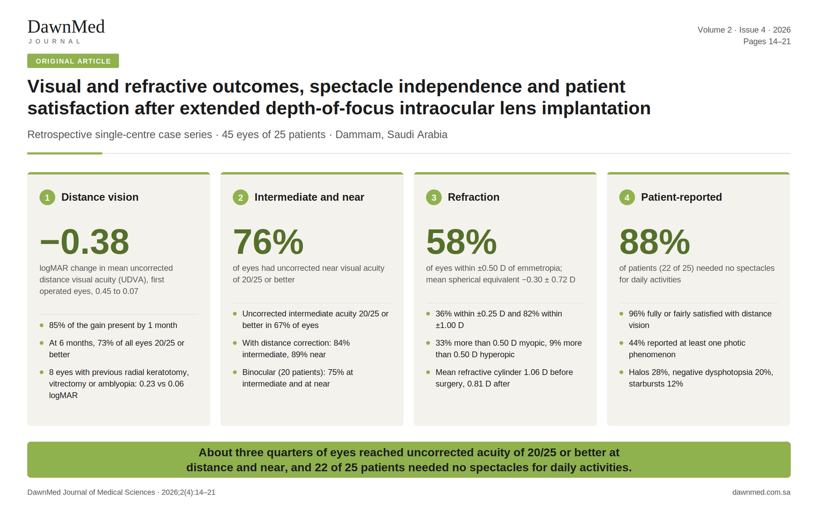
Results: In first eyes, mean UDVA improved from 0.45 ± 0.23 to 0.07 ± 0.09 logMAR (change −0.38, 95% confidence interval [CI] −0.47 to −0.28; p < 0.001), and 85% of this change had occurred by 1 month. At 6 months, 73% of all eyes had UDVA of 20/25 or better. Uncorrected intermediate and near acuity were 20/25 or better in 67% and 76% of eyes (84% and 89% with distance correction). Postoperative spherical equivalent was −0.30 ± 0.72 D and was within ±0.50 D of emmetropia in 58% of eyes. Twenty-two of 25 patients (88%) reported needing no spectacles for daily activities, and 24 (96%) were fully or fairly satisfied with distance vision. Eleven patients (44%) reported at least one photic phenomenon, most often halos (28%). The eight eyes with previous radial keratotomy, vitrectomy or amblyopia had a mean 6-month UDVA of 0.23 logMAR, compared with 0.06 logMAR in the other 37 eyes.
Conclusions: After EDOF IOL implantation, about three quarters of eyes reached uncorrected acuity of 20/25 or better at distance and near, and most patients reported not needing spectacles. Photic phenomena were reported by 44% of patients. Without a control group, these outcomes cannot be attributed to the lens designs.
Introduction
Extended depth-of-focus (EDOF) intraocular lenses (IOLs) use optical designs that increase the depth of focus of the pseudophakic eye, with the aim of improving intermediate vision while having little effect on distance vision [1]. The American Academy of Ophthalmology task force defined the category by three criteria: monocular corrected distance acuity comparable to that of a monofocal control, tested for non-inferiority; a monocular depth of focus at least 0.50 D greater than that of the monofocal control at 0.2 logMAR (logarithm of the minimum angle of resolution); and mean monocular distance-corrected intermediate acuity at 66 cm superior to that of the control, with at least 50% of eyes reaching 0.2 logMAR or better [1]. Meta-analyses comparing multifocal with monofocal IOLs have found greater spectacle independence with multifocal lenses, at the cost of more glare and halos [2,3]. In a non-randomised comparison, an extended range of vision IOL gave better uncorrected distance, intermediate and near acuity than an aspheric monofocal IOL [4].
In the multicentre Concerto study, 411 patients had bilateral implantation of an extended range of vision IOL, and more than 90% reported no or only mild halos, glare, starbursts or other photic phenomena 4 to 6 months after surgery [5]. Some candidates for presbyopia-correcting lenses have had previous corneal or retinal surgery or have reduced visual potential, and the American Society of Cataract and Refractive Surgery (ASCRS) Cataract Clinical Committee has published guidance on patient selection for multifocal IOLs that addresses eyes with coexisting ocular conditions [6]. In a study of dissatisfied patients with multifocal IOLs, the most common identified causes of blurred vision were posterior capsule opacification, residual refractive error and dry eye [7].
We report uncorrected distance acuity over the first 6 months, and postoperative intermediate and near acuity, refraction, spectacle use, photic phenomena and satisfaction, in a series of eyes that received an EDOF IOL at one private centre in Saudi Arabia. The series included a small number of eyes with previous radial keratotomy, pars plana vitrectomy or amblyopia, which are described separately.
Methods
Design and setting
This retrospective single-centre case series reviewed the clinical records of patients who underwent cataract surgery with implantation of an EDOF IOL at Al Kahhal Medical Complex, Dammam, Saudi Arabia, between August 2022 and September 2023. Distance acuity was recorded up to 6 months after each operation. Patient data were de-identified before analysis. The study followed the Declaration of Helsinki, and written informed consent was obtained from all patients; the ethics statement is given under Declarations.
Patients
The study included 45 eyes of 25 adults who underwent phacoemulsification with EDOF IOL implantation. Both eyes of a patient were included when both were operated. Eyes with previous radial keratotomy, previous pars plana vitrectomy or amblyopia are described separately as complex eyes.
Preoperative assessment and IOL power
Before surgery, uncorrected (UDVA) and corrected distance visual acuity (CDVA) were measured with a Snellen chart and recorded in decimal notation, and subjective refraction was performed. Corneal topography was assessed with a Scheimpflug imaging device, and anterior and posterior segment findings were recorded. The biometry method and the intended refraction were not recorded in the clinical records reviewed for this study.
Surgical technique
One surgeon performed all operations under topical anaesthesia. Through a 2.2 mm clear corneal incision, the cataract was removed by phacoemulsification, cortical material was removed, and one of two EDOF IOLs, the Bi-Flex ELON (Medicontur Medical Engineering, Zsámbék, Hungary) or the Synthesis+ (Cutting Edge, Montpellier, France), was implanted in the capsular bag with an injector. The lens model was not recorded for individual eyes. In the 20 patients who had both eyes operated, the second eye was operated on a median of 1 day after the first (range 0 to 173 days).
Outcome measures
UDVA was recorded at 2 weeks and at 1, 3 and 6 months after surgery. At one further postoperative assessment, the following were recorded for each eye: monocular UDVA, uncorrected intermediate (UIVA) and near (UNVA) visual acuity, and distance-corrected intermediate (DCIVA) and near (DCNVA) visual acuity; binocular UDVA, CDVA, UIVA, DCIVA, UNVA and DCNVA; and refraction. The same records contain patients’ answers to a questionnaire in which they rated their satisfaction with distance, intermediate and near vision separately as fully satisfied, fairly satisfied or not satisfied, stated whether they needed spectacles for any daily activity, and stated whether they noticed starbursts, halos or negative dysphotopsia. The clinical records did not specify the visit at which these measurements and the questionnaire were recorded, the test distances and charts used for intermediate and near acuity, or whether postoperative refraction was subjective or automated.
Statistical analysis
Decimal acuities were converted to logMAR, and Snellen equivalents of mean values are given for orientation. Because both eyes of 20 patients were included, inferential analyses used one eye per patient: the first operated eye, identified by surgery date, with the right eye taken when both eyes were operated on the same day (one patient). This rule was defined after data collection and was applied without reference to outcomes. Results for all 45 eyes are presented as descriptive summaries, without confidence intervals or p-values. Paired comparisons in first eyes used the paired t-test with a 95% confidence interval (CI) for the mean change; the Wilcoxon signed-rank test was run alongside, and both p-values are reported. Paired differences were checked for normality with the Shapiro–Wilk test. The four postoperative UDVA measurements were compared with the Friedman test. Proportions in first eyes and in patients are given with 95% Wilson CIs.
Binocular acuity is reported for the 20 bilaterally implanted patients only, because the status of the fellow eye in unilateral patients was not recorded. In three bilateral patients the binocular values differed between the records of the two eyes; the values recorded with the later-operated eye were used, and the effect of using the other record is reported. Questionnaire outcomes were summarised once per patient using the least favourable response recorded for either eye; a patient was classed as spectacle independent only if the records of both eyes said so, and as having a photic phenomenon if the record of either eye did. Refraction is expressed relative to emmetropia because the intended refraction was not recorded.
Comparisons between subgroups were exploratory and used the Fisher exact test, the Mann–Whitney U test and Spearman correlation at patient or first-eye level; comparisons involving the 8 complex eyes are descriptive only. Two preoperative refraction entries were ambiguous: a cylinder recorded without a sign, analysed as −0.50 D in keeping with the minus-cylinder notation of all other records, and a sphere recorded as “+225”, analysed as +2.25 D. Sensitivity analyses report the effect of excluding the second eye and of reading the first cylinder as +0.50 D. One monocular UDVA entry at the postoperative assessment was recorded as 0 and was treated as missing. No sample size calculation was performed. Analyses were performed in Python 3 with the SciPy library.
Results
Patients
Forty-five eyes of 25 patients were included; 20 patients had both eyes operated and 5 had one eye operated. Eleven patients (44%) were men. Mean age was 53.4 ± 9.6 years (range 26 to 70). Thirty-seven eyes (82%) had no previous ocular surgery or amblyopia recorded. Four eyes of two patients had previous radial keratotomy, three eyes of two patients had previous pars plana vitrectomy (both eyes of one patient for retinal detachment and one eye of another for a macular hole), and one eye was amblyopic. Recorded posterior segment findings included a tigroid fundus in six eyes of three patients, four of them eyes with previous radial keratotomy, and posterior vitreous detachment in both eyes of one patient. Preoperative spherical equivalent ranged from −5.88 to +3.50 D; 29 eyes were myopic, 1 was plano and 15 were hyperopic. Before surgery, 12 eyes had CDVA of 20/40 or worse and 10 had CDVA of 20/20. All 45 eyes had UDVA recorded at 6 months (Table 1).
Table 1. Baseline characteristics of the 45 eyes (descriptive).
Characteristic
Value
Patients / eyes, n
25 / 45
Bilateral / unilateral patients, n
20 / 5
Age, years, mean ± SD (range)
53.4 ± 9.6 (26–70)
Sex, male / female, n (%)
11 (44) / 14 (56)
Eyes without previous ocular surgery or amblyopia, n (%)
Spherical equivalent, D, mean ± SD (median; range)
−1.05 ± 2.52 (−1.63; −5.88 to +3.50)
Myopic / plano / hyperopic eyes, n
29 / 1 / 15
Refractive cylinder, D, mean ± SD (median)
1.06 ± 0.82 (1.00)
Interval between eyes in bilateral patients, days, median (range)
1 (0–173)
CDVA, corrected distance visual acuity; D, dioptre; SD, standard deviation; UDVA, uncorrected distance visual acuity. Preoperative refraction includes one sphere recorded as “+225” (analysed as +2.25 D) and one cylinder recorded without a sign (analysed as −0.50 D); sensitivity analyses are given under Refractive outcomes.
Uncorrected distance acuity over time
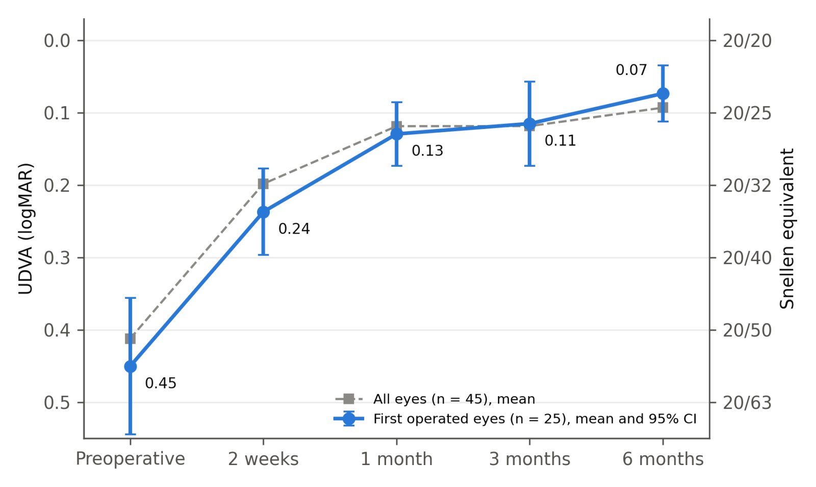
In first eyes, UDVA improved from 0.45 ± 0.23 logMAR before surgery to 0.24 ± 0.15 at 2 weeks, 0.13 ± 0.11 at 1 month, 0.11 ± 0.14 at 3 months and 0.07 ± 0.09 at 6 months (Friedman χ² = 40.2 across the four postoperative visits, p < 0.001; Table 2, Figure 1). The mean change from before surgery to 6 months was −0.38 logMAR (95% CI −0.47 to −0.28; paired t-test p < 0.001, Wilcoxon p < 0.001), a gain of almost four lines. Acuity improved between 2 weeks and 1 month (mean change −0.11, 95% CI −0.15 to −0.07; p < 0.001), did not change significantly between 1 and 3 months (−0.01, 95% CI −0.07 to 0.04; p = 0.606, Wilcoxon p = 0.244), and improved by a further 0.04 logMAR between 3 and 6 months (95% CI −0.07 to −0.01; p = 0.011, Wilcoxon p = 0.005). By 1 month, 85% of the total change had occurred. At 6 months, 21 of 25 first eyes and 35 of 45 eyes overall had UDVA equal to or better than their preoperative CDVA. Between 1 and 6 months, UDVA fell by 0.2 logMAR or more in three eyes: both eyes of a 70-year-old man and the eye that had undergone vitrectomy for a macular hole. The cause was not recorded.
Table 2. Monocular uncorrected distance visual acuity (logMAR) by visit.
Visit
First eyes (n = 25), mean ± SD
Snellen equivalent
Change from previous visit (95% CI)
p (paired t; Wilcoxon)
All eyes (n = 45), mean ± SD
Preoperative
0.45 ± 0.23
20/56
0.41 ± 0.23
2 weeks
0.24 ± 0.15
20/35
−0.21 (−0.31 to −0.12)
< 0.001; < 0.001
0.20 ± 0.14
1 month
0.13 ± 0.11
20/27
−0.11 (−0.15 to −0.07)
< 0.001; < 0.001
0.12 ± 0.10
3 months
0.11 ± 0.14
20/26
−0.01 (−0.07 to 0.04)
0.606; 0.244
0.12 ± 0.14
6 months
0.07 ± 0.09
20/24
−0.04 (−0.07 to −0.01)
0.011; 0.005
0.09 ± 0.16
Inference is based on the first operated eye of each patient; all-eye values are descriptive. Snellen equivalents refer to first-eye means. Overall change in first eyes from before surgery to 6 months: −0.38, 95% CI −0.47 to −0.28; p < 0.001.
Figure 1. Mean monocular uncorrected distance visual acuity (UDVA) from before surgery to 6 months. Circles: first operated eye of each patient (n = 25), with 95% confidence intervals. Squares: all 45 eyes, descriptive means.
Acuity at distance, intermediate and near
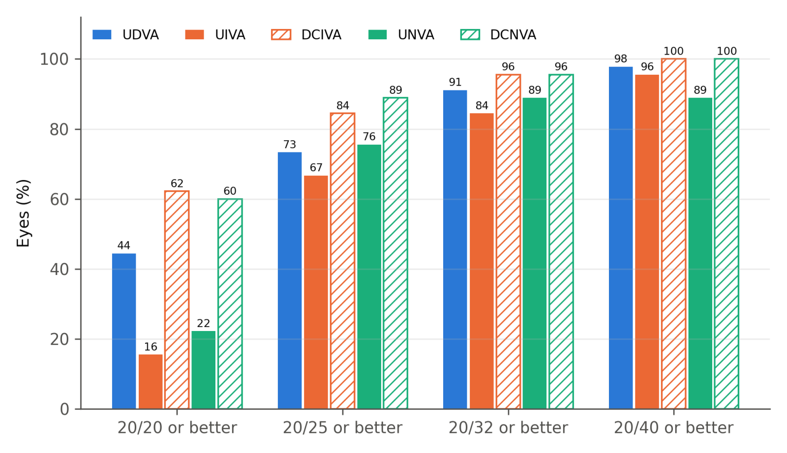
Table 3 and Figure 2 give acuity at each distance. At 6 months, 73% of all eyes had UDVA of 20/25 or better and 44% had 20/20 or better; 91% reached 20/32 and 98% reached 20/40. At the postoperative assessment, UIVA was 20/25 or better in 67% of eyes and UNVA in 76%; with distance correction, 84% reached 20/25 or better at intermediate and 89% at near, and no eye read worse than 20/40 at either distance. In first eyes, the proportions with 20/25 or better were 76% (95% CI 57–89) for UDVA, 64% (95% CI 45–80) for UIVA and 76% (95% CI 57–89) for UNVA. The monocular UDVA recorded at the postoperative assessment was identical to the 6-month value in 23 of 44 evaluable eyes and within 0.1 logMAR in 39.
Among the 20 bilaterally implanted patients, binocular uncorrected acuity was 20/25 or better in 15 (75%, 95% CI 53–89) at distance, 15 at intermediate and 15 at near, and binocular DCNVA was 20/25 or better in all 20. Using the other eye’s record for the three patients with discrepant binocular values changed two results by one patient each: binocular UDVA of 20/20 or better in 10 rather than 11 patients, and binocular UNVA of 20/25 or better in 16 rather than 15.
UDVA is from the 6-month visit; the other monocular values and all binocular values are from the postoperative assessment described in Methods. For binocular values, the record of the later-operated eye was used. CDVA, corrected distance visual acuity; DCIVA/DCNVA, distance-corrected intermediate/near visual acuity; UDVA, uncorrected distance visual acuity; UIVA/UNVA, uncorrected intermediate/near visual acuity.
Figure 2. Cumulative proportion of all 45 eyes reaching each Snellen level (descriptive). UDVA is from the 6-month visit; intermediate and near values are from the postoperative assessment. Hatched bars show distance-corrected acuity.
Refractive outcomes
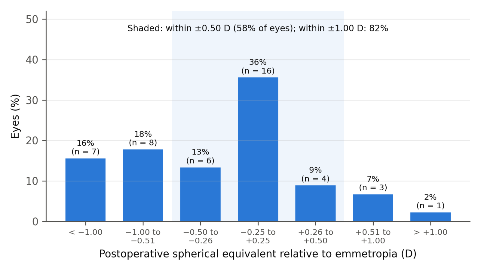
Across all 45 eyes, postoperative spherical equivalent was −0.30 ± 0.72 D (median −0.25 D; Table 4, Figure 3). Sixteen eyes (36%) were within ±0.25 D of emmetropia, 26 (58%) within ±0.50 D and 37 (82%) within ±1.00 D; 15 eyes (33%) were more than 0.50 D myopic and four (9%) more than 0.50 D hyperopic. Refractive cylinder was 0.81 ± 0.43 D, and eight eyes (18%) had more than 1.00 D. In first eyes, absolute spherical equivalent fell from 2.45 ± 1.40 D to 0.64 ± 0.53 D (mean change −1.82 D, 95% CI −2.39 to −1.24; p < 0.001); 14 of 25 (56%, 95% CI 37–73) were within ±0.50 D and 21 (84%, 95% CI 65–94) within ±1.00 D. Refractive cylinder in first eyes fell from 1.22 ± 0.93 D to 0.89 ± 0.37 D (mean change −0.33 D, 95% CI −0.66 to 0.00; p = 0.053, Wilcoxon p = 0.056).
Both eyes with an ambiguous preoperative entry were first eyes, so the entries affect the preoperative summaries in Table 1 and the paired first-eye comparisons in Table 4; their postoperative refraction and acuity values are unambiguous. Excluding the eye recorded as “+225” gave a mean preoperative spherical equivalent of −1.12 ± 2.50 D across 44 eyes (14 hyperopic) and, in 24 first eyes, a mean change in absolute spherical equivalent of −1.83 D (95% CI −2.43 to −1.22) and in cylinder of −0.34 D (95% CI −0.69 to 0.00; p = 0.052, Wilcoxon p = 0.056). Reading the unsigned cylinder as +0.50 D gave a mean preoperative spherical equivalent of −1.04 D and a first-eye change in absolute spherical equivalent of −1.80 D (95% CI −2.36 to −1.23).
Table 4. Refractive outcomes.
Measure
Preoperative
Postoperative
p (paired t; Wilcoxon)
All eyes, descriptive (n = 45)
Spherical equivalent, D, mean ± SD (median)
−1.05 ± 2.52 (−1.63)
−0.30 ± 0.72 (−0.25)
Within ±0.25 / ±0.50 / ±1.00 D of emmetropia, n (%)
16 (36) / 26 (58) / 37 (82)
More than 0.50 D myopic / hyperopic, n (%)
15 (33) / 4 (9)
Refractive cylinder, D, mean ± SD (median)
1.06 ± 0.82 (1.00)
0.81 ± 0.43 (0.75)
Cylinder ≤ 0.50 / ≤ 1.00 D, n (%)
16 (36) / 37 (82)
First eyes (n = 25)
Absolute spherical equivalent, D, mean ± SD
2.45 ± 1.40
0.64 ± 0.53
< 0.001; < 0.001
Within ±0.50 D of emmetropia, % (95% CI)
56 (37–73)
Within ±1.00 D of emmetropia, % (95% CI)
84 (65–94)
Refractive cylinder, D, mean ± SD
1.22 ± 0.93
0.89 ± 0.37
0.053; 0.056
Postoperative refraction is from the postoperative assessment described in Methods. Spherical equivalent is expressed relative to emmetropia because the intended refraction was not recorded. D, dioptre; SD, standard deviation.
Figure 3. Distribution of postoperative spherical equivalent relative to emmetropia (all 45 eyes). The shaded categories are within ±0.50 D.
Satisfaction, spectacle use and photic phenomena
Twenty-two of 25 patients (88%, 95% CI 70–96) reported needing no spectacles for any daily activity; the records of the two eyes agreed in all 20 bilateral patients. Using each patient’s least favourable response, 17 patients (68%) were fully satisfied with distance vision, seven (28%) fairly satisfied and one (4%) not satisfied; the corresponding numbers were 20, 4 and 1 for intermediate vision and 20, 3 and 2 for near vision (Table 5). The records of the two eyes gave the same distance rating in 17 of the 20 bilateral patients. Fourteen patients (56%) were fully satisfied at all three distances. Eleven patients (44%, 95% CI 27–63) reported at least one photic phenomenon in either eye: halos in seven (28%), negative dysphotopsia in five (20%) and starbursts in three (12%). The patient who was not satisfied at any distance was the 70-year-old man whose UDVA fell in both eyes after the first month; he reported all three phenomena in both eyes and needed spectacles.
Table 5. Patient-reported outcomes.
Measure
Fully satisfied, n (%)
Fairly satisfied, n (%)
Not satisfied, n (%)
Fully or fairly satisfied, % (95% CI)
Satisfaction per patient, least favourable eye record (n = 25)
Distance vision
17 (68)
7 (28)
1 (4)
96 (80–99)
Intermediate vision
20 (80)
4 (16)
1 (4)
96 (80–99)
Near vision
20 (80)
3 (12)
2 (8)
92 (75–98)
Other patient-level outcomes (n = 25)
Patients, n
% (95% CI)
Eye records, n/45
No spectacles needed for any daily activity
22
88 (70–96)
40/45
Any photic phenomenon (either eye)
11
44 (27–63)
19/45
Halos
7
28 (14–48)
13/45
Negative dysphotopsia
5
20 (9–39)
8/45
Starbursts
3
12 (4–30)
5/45
Patient-level values use the least favourable response recorded for either eye; counts of eye records are shown for reference and are not independent observations.
Exploratory comparisons
Six of 11 patients with a photic phenomenon were fully satisfied with distance vision, compared with 11 of 14 without (p = 0.389; Table 6). In first eyes, mean UDVA was 0.12 logMAR in the 11 patients with a photic phenomenon and 0.03 logMAR in the 14 without (p = 0.074), and mean absolute spherical equivalent was 0.67 D and 0.61 D (p = 0.679). Among first eyes, 12 of 14 within ±0.50 D of emmetropia had UDVA of 20/25 or better, compared with 7 of 11 outside this range (p = 0.350), and the correlation between absolute spherical equivalent and UDVA was weak (Spearman ρ = −0.03, p = 0.885).
The eight complex eyes, from five patients, had a mean 6-month UDVA of 0.23 ± 0.32 logMAR, compared with 0.06 ± 0.09 logMAR in the other 37 eyes; 4 of 8 reached 20/25 or better, compared with 29 of 37 (78%). Only four complex eyes were first eyes, so these groups were not compared statistically. All four eyes with previous radial keratotomy reached 20/25 or better, each had at least one photic phenomenon recorded, and three of the four were rated fairly rather than fully satisfied for distance. The three eyes with previous vitrectomy reached 20/32, 20/29 and 20/200, and the amblyopic eye reached 20/29.
Table 6. Exploratory comparisons.
Comparison
Group 1
Group 2
p
Fully satisfied with distance vision: patients with vs without a photic phenomenon
6/11 (55%)
11/14 (79%)
0.389 (Fisher)
UDVA at 6 months, logMAR, first eyes: patients with vs without a photic phenomenon
0.12 (n = 11)
0.03 (n = 14)
0.074 (Mann–Whitney)
Absolute spherical equivalent, D, first eyes: with vs without a photic phenomenon
0.67
0.61
0.679 (Mann–Whitney)
UDVA 20/25 or better, first eyes: within vs outside ±0.50 D of emmetropia
12/14 (86%)
7/11 (64%)
0.350 (Fisher)
UDVA at 6 months, logMAR, all eyes: complex vs other eyes
0.23 ± 0.32 (n = 8)
0.06 ± 0.09 (n = 37)
not tested
UDVA 20/25 or better, all eyes: complex vs other eyes
4/8
29/37 (78%)
not tested
Tests were applied only at patient or first-eye level; comparisons of complex with other eyes are descriptive because the eight complex eyes came from five patients. UDVA, uncorrected distance visual acuity.
Discussion
In this series, UDVA in first eyes improved by a mean of 0.38 logMAR, mostly within the first month, and about three quarters of eyes had uncorrected acuity of 20/25 or better at distance and near. Twenty-two of 25 patients reported needing no spectacles for daily activities. Among the 20 bilaterally implanted patients, binocular uncorrected acuity was 20/25 or better in 15 at each of the three distances.
Photic phenomena were reported by 44% of patients. Differences in questionnaire definitions and severity grading prevent direct comparison with published trials. Our questionnaire recorded whether each phenomenon was present, without grading its severity or how much it bothered the patient, whereas the Concerto study reported the proportion of patients with no or only mild phenomena [5]. All four eyes with previous radial keratotomy in this series were recorded as having at least one photic phenomenon.
Postoperative spherical equivalent was within ±0.50 D of emmetropia in 58% of eyes and within ±1.00 D in 82%, with a mean of −0.30 D, and 18% of eyes had more than 1.00 D of refractive cylinder. Because the intended refraction was not recorded, these figures describe refraction relative to emmetropia and not the accuracy of the IOL power calculation; eyes deliberately targeted for mild myopia would appear here as refractive error. Son et al. found that acuity with an EDOF IOL was similar to that with a monofocal IOL on the same platform within ±0.50 D of defocus and better at greater defocus [8]. In our first eyes, the correlation between residual spherical equivalent and UDVA was weak, and 7 of 11 eyes outside ±0.50 D still reached 20/25. In an analysis of retreatments after multifocal IOL implantation, complaints related to residual refractive error were the main reason for retreatment, and retreated eyes had more residual astigmatism [9]. The reduction in cylinder in our first eyes did not reach significance.
Full satisfaction with distance vision was less frequent among patients who reported photic phenomena (6 of 11) than among those who did not (11 of 14), but the difference was not significant, and with 25 patients a difference of clinically relevant size cannot be excluded. First eyes of patients with photic phenomena also had lower mean UDVA without a difference in residual refraction, and this difference was not significant either. Woodward et al. identified posterior capsule opacification, residual refractive error and dry eye as the most common causes of blurred vision in dissatisfied patients with multifocal IOLs [7]. Ocular surface status and posterior capsule opacification were not recorded in our series, and the patient who was dissatisfied at every distance lost acuity in both eyes after the first month for reasons that were not recorded.
The eight complex eyes are described rather than tested because they came from five patients and only four were first eyes. Their mean UDVA was lower than that of the other eyes. The four eyes with previous radial keratotomy all reached 20/25 or better, but each was recorded as having a photic phenomenon, and three of the four were rated fairly rather than fully satisfied for distance. Before surgery, CDVA in the three vitrectomised eyes and the amblyopic eye ranged from 20/80 to 20/29, and the eye with a previous macular hole reached 20/200 at 6 months. The ASCRS guidance on multifocal IOLs addresses patient selection in eyes with coexisting ocular conditions [6]; with so few eyes, our data do not allow conclusions about EDOF lenses in these groups.
Most of the improvement in UDVA had occurred by 1 month, with a further small gain of 0.04 logMAR between 3 and 6 months. Uncorrected near acuity was slightly better than intermediate acuity in this series. Kohnen et al. reported better binocular uncorrected intermediate acuity at 60 and 80 cm than near acuity with an EDOF IOL [10]; without the test distances used here, the two sets of results cannot be compared.
The series has UDVA at four postoperative visits for every eye, acuity at three distances measured monocularly and binocularly with and without distance correction, and photic phenomena for each eye, and the inferential analyses account for the inclusion of fellow eyes.
Limitations
This was a single-centre series of 25 patients without a control group, and the estimates are imprecise: the proportion of first eyes reaching 20/25 uncorrected, for example, has a 95% CI of 57–89%. Without a comparison group, the outcomes cannot be attributed to the EDOF optics rather than to cataract surgery itself, and they give no basis for comparing these lenses with monofocal or multifocal alternatives. Two EDOF designs were used and the lens model was not recorded for individual eyes, so the results describe the two lenses together and cannot be assigned to either.
Both eyes of 20 patients were included. Inferential tests used one eye per patient, which removes the dependence between fellow eyes but reduces the sample available for testing to 25, and the eye-selection rule was defined after data collection. The all-eye summaries are descriptive and would overstate precision if read as 45 independent observations. The complex-eye subgroup contained only eight eyes with three different conditions, and all subgroup comparisons were exploratory; with groups this small, the absence of a significant difference is compatible with a clinically important one.
As a retrospective series, the study is open to selection bias. All included eyes had records to 6 months, and patients who received these lenses during the study period but were not included are not described, so the series may not represent all patients treated.
Satisfaction was recorded in three categories and photic phenomena as present or absent, using a questionnaire without published validation or grading of severity. These measures show whether a symptom occurred but not how much it affected the patient. Intermediate, near and binocular acuity and postoperative refraction were recorded at a single postoperative assessment, so their time course is unknown; the test distances were not recorded, which limits comparison with other studies; and in three patients the records of the two eyes carried different binocular values, although the choice between them changed only two threshold results by one patient each.
The intended refraction was not recorded, so the refractive results describe distance from emmetropia rather than prediction error, and eyes deliberately left slightly myopic appear less accurate than they may have been. Defocus curves, contrast sensitivity and aberrometry were not analysed, so the depth of focus and optical quality of the lenses could not be assessed directly. Follow-up ended at 6 months, and the reasons for the loss of acuity after the first month in three eyes were not recorded.
Conclusion
In this series, EDOF IOL implantation was followed by uncorrected acuity of 20/25 or better at distance and near in about three quarters of eyes and by reported spectacle independence in 22 of 25 patients, with most of the gain in distance acuity present by 1 month. Photic phenomena were reported by 44% of patients, and postoperative spherical equivalent was within ±0.50 D of emmetropia in 58% of eyes. Outcomes in the few eyes with previous ocular surgery or amblyopia are reported descriptively.
Declarations
Ethics statement: This retrospective study used de-identified clinical data. Al Kahhal Medical Complex does not have an institutional review board; the management of Al Kahhal Medical Complex reviewed the study and granted institutional authorisation (letter of the Medical Director, 10 September 2026). The study followed the Declaration of Helsinki and the guidelines of the Saudi National Committee of Bioethics.
Consent to participate: Written informed consent was obtained from all patients.
Funding: This research received no specific grant from any funding agency in the public, commercial or not-for-profit sectors.
Conflicts of interest: The authors declare that they have no conflicts of interest.
Data availability: The de-identified per-eye dataset is available from the corresponding author on reasonable request.
Author contributions: All authors contributed equally to this work and approved the final manuscript.
Use of artificial intelligence: AI-assisted tools (Claude, Anthropic) were used during preparation of the revised manuscript for language editing, re-running the statistical analysis from the source data, and formatting of tables, figures and references. All AI-assisted output was checked against the source data and reviewed by the authors, who approved the final text and take full responsibility for its content. The editorial office used AI tools for the initial screening of the submission; all editorial and peer-review decisions were made by the editors and reviewers.
References
MacRae S, Holladay JT, Glasser A, et al. Special report: American Academy of Ophthalmology Task Force consensus statement for extended depth of focus intraocular lenses. Ophthalmology. 2017;124(1):139-141. doi:10.1016/j.ophtha.2016.09.039
Khandelwal SS, Jun JJ, Mak S, Booth MS, Shekelle PG. Effectiveness of multifocal and monofocal intraocular lenses for cataract surgery and lens replacement: a systematic review and meta-analysis. Graefes Arch Clin Exp Ophthalmol. 2019;257(5):863-875. doi:10.1007/s00417-018-04218-6
Cao K, Friedman DS, Jin S, et al. Multifocal versus monofocal intraocular lenses for age-related cataract patients: a system review and meta-analysis based on randomized controlled trials. Surv Ophthalmol. 2019;64(5):647-658. doi:10.1016/j.survophthal.2019.02.012
Pedrotti E, Bruni E, Bonacci E, Badalamenti R, Mastropasqua R, Marchini G. Comparative analysis of the clinical outcomes with a monofocal and an extended range of vision intraocular lens. J Refract Surg. 2016;32(7):436-442. doi:10.3928/1081597X-20160428-06
Cochener B; Concerto Study Group. Clinical outcomes of a new extended range of vision intraocular lens: International Multicenter Concerto Study. J Cataract Refract Surg. 2016;42(9):1268-1275. doi:10.1016/j.jcrs.2016.06.033
Braga-Mele R, Chang D, Dewey S, et al.; ASCRS Cataract Clinical Committee. Multifocal intraocular lenses: relative indications and contraindications for implantation. J Cataract Refract Surg. 2014;40(2):313-322. doi:10.1016/j.jcrs.2013.12.011
Son HS, Kim SH, Auffarth GU, Choi CY. Prospective comparative study of tolerance to refractive errors after implantation of extended depth of focus and monofocal intraocular lenses with identical aspheric platform in Korean population. BMC Ophthalmol. 2019;19(1):187. doi:10.1186/s12886-019-1193-z
Gundersen KG, Makari S, Ostenstad S, Potvin R. Retreatments after multifocal intraocular lens implantation: an analysis. Clin Ophthalmol. 2016;10:365-371. doi:10.2147/OPTH.S100840
Kohnen T, Böhm M, Hemkeppler E, et al. Visual performance of an extended depth of focus intraocular lens for treatment selection. Eye (Lond). 2019;33(10):1556-1563. doi:10.1038/s41433-019-0443-x